
Volume 3, Issue 4, 2025
Editorial
Reframing the Approach to Predatory Journals; Embracing a 'Non-Recommended Journal' Model
Abdullah Khalid Omer
For more than a decade, the academic publishing community has been locked in a battle against “predatory journals.” These are commonly understood as outlets that exploit the open access model by charging fees to authors without providing genuine peer review or editorial services [1]. While this campaign has been well-intentioned, its implementation has been riddled with inconsistencies and collateral damage. It is time to re-evaluate our approach—and a promising alternative has recently been proposed.
At the 18th Meeting of the European Association of Science Editors (EASE), Kakamad et al. introduced the concept of the Non-Recommended Journal (NRJ), offering a more nuanced and constructive way to classify questionable journals. Their proposal, outlined in a poster presented at the event, acknowledges a critical truth that the current binary model overlooks: not all low-quality or problematic journals are predatory, and not all accused journals are guilty [2].
One of the core issues with the term “predatory” is its lack of a universally accepted definition. Attempts to label journals as predatory can often be subjective and based on flawed or incomplete criteria. This ambiguity has led to wrongful accusations and the potential defamation of emerging or under-resourced journals that are making genuine efforts to improve. Worse still, some well-established journals exhibit questionable practices yet avoid scrutiny simply because they don’t fit the “predatory” mold [3].
The NRJ framework reframes the discussion by focusing not on intention, but on recommendation. Rather than asking whether a journal is maliciously exploitative, the NRJ model asks whether a journal meets acceptable standards of transparency, editorial rigor, and academic integrity. Journals that do not meet these standards—whether due to deliberate misconduct or lack of infrastructure can be flagged as “non-recommended” without implying criminality or predation [2].
This shift in terminology allows for a more flexible and inclusive way to monitor journal quality. It accounts for the so-called “borderline journals,” which may not be outright deceptive but still fail to uphold scholarly standards. By avoiding the inflammatory label of “predatory,” the NRJ system reduces the risk of reputational harm while still guiding authors, reviewers, and institutions toward better publishing decisions.
Moreover, the NRJ approach invites continuous re-evaluation. Journals can move in and out of this category based on demonstrated improvements, providing a growth mindset rather than cementing stigmas. This dynamic classification also encourages more transparent criteria, ideally informed by independent watchdogs or academic associations rather than commercial blacklists.
It is time we recognize the complexity of the academic publishing ecosystem and evolve beyond the simplistic predator-prey narrative. The NRJ concept represents a practical, fair, and forward-thinking step in that direction. As the academic world continues to grapple with questions of quality, ethics, and accessibility, such innovations are not just welcome, they are essential.
Original Articles

Predictors of False-Negative Axillary FNA Among Breast Cancer Patients: A Cross-Sectional Study
Lana R. A. Pshtiwan, Sakar O. Arif, Harzal Hiwa Fatih, Masty K. Ahmed, Shaban Latif, Meer M....
Abstract
Introduction
Fine-needle aspiration (FNA) is commonly used to investigate lymphadenopathy of suspected metastatic origin. The current study aims to find the association between nodal characteristics and cancer-related factors with the rate of false-negative preoperative FNA.
Methods
This retrospective, single-center, cross-sectional study included breast cancer patients with negative preoperative axillary FNA results who underwent postoperative histopathological evaluation. Data were collected from electronic medical records, including clinical, imaging, cytological, and pathological findings. Patients with incomplete records, non-axillary or inconclusive FNAs, positive preoperative FNAs, or unsampled axillae postoperatively were excluded. Key variables analyzed included lymph node size, cortical thickness, tumor grade, histological type, immunohistochemical subtype, and metastatic patterns.
Results
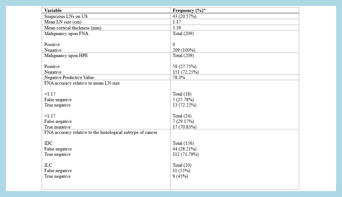
A total of 209 negative axillary FNA samples were analyzed, with a mean patient age of 46.13 years. Invasive ductal carcinoma was the most common diagnosis, and ER-positive tumors were the predominant subtype. Ultrasonography identified suspicious axillary nodes in 20.57% of cases. Histopathology revealed a 27.75% false-negative rate, with a negative predictive value of 78.3%. Larger lymph node size and cortical thickness exhibited lower false-negative rates, while histologic type and ER status showed significant associations with false-negative outcomes (P < 0.05).
Conclusion
The 27.75% false-negative rate of preoperative FNA remains concerning and may not be sufficiently low to justify foregoing definitive axillary staging. The current study found significant associations between false-negative FNA rates and histological subtype and ER status, the latter of which is not explicitly mentioned in the literature.
Introduction
Breast cancer represents the most commonly diagnosed malignancy among women globally. In 2020, it surpassed lung cancer to become the leading cancer diagnosis in women worldwide, with an estimated 2.3 million new cases reported [1,2]. Precise clinical staging plays a pivotal role in assessing prognosis, informing treatment strategies, and anticipating clinical outcomes in breast cancer. A critical component of staging is the evaluation of axillary lymph node involvement, as the presence or absence of metastasis in these nodes substantially influences therapeutic approaches and overall survival rates [3].
Conventional techniques, including sentinel lymph node biopsy (SLNB) and axillary lymph node dissection (ALND), have long been the standard approaches for axillary staging in newly diagnosed breast cancer patients. Despite their diagnostic utility, these invasive procedures are associated with inherent risks and potential postoperative complications, notably lymphedema [1].
Axillary lymph node fine-needle aspiration biopsy (FNAB) has gained recognition as an effective method for staging breast cancer. When performed under ultrasound guidance (US-FNA), this technique allows for targeted sampling of suspicious axillary lymph nodes through a minimally invasive approach, providing a less invasive and potentially less morbid alternative to traditional methods such as SLNB and ALND [4]. This technique exhibits high diagnostic accuracy in detecting metastatic disease. Although the overall diagnostic accuracy of ultrasound-guided FNAB is well documented, the understanding of lymph node characteristics that influence the technique's sensitivity for detecting metastatic disease is limited [5]. Ewing et al. demonstrated that true-positive FNAB results were more likely in larger lymph nodes compared to false-negative results. Furthermore, true positive FNABs were associated with a higher percentage of nodal replacement by carcinoma, in contrast to false negative FNABs [6]. Establishing a precise correlation between the diagnostic sensitivity of FNAB for detecting metastatic malignancies and specific nodal characteristics remains challenging. The current study aims to establish the relationship between nodal characteristics and cancer-related factors with the false-negative FNAB rate through a retrospective analysis of data from 209 patients.
Methods
Study design
This was a retrospective, single-center, cross-sectional study. Data were collected over two months, from November to December 2024. All participants provided informed consent, including agreement to the use of their anonymized data for publication purposes. This study was approved by the Ethics Committee of the Kscien organization (Approval No. 2025-34).
Data source
Data were obtained through a review of electronic medical records, which included demographic information, medical history, presenting complaints, preoperative imaging findings (ultrasound, magnetic resonance imaging, and computed tomography), FNA results, diagnosis, treatment approaches, and postoperative histopathological outcomes.
Eligibility criteria
The study included patients diagnosed with breast cancer who had negative axillary FNA results before surgery and underwent histopathological assessment postoperatively. Exclusion criteria included patients with incomplete medical records, FNAs not sourced from the axillary region, positive preoperative axillary FNA results, FNAs conducted outside the study center, inconclusive FNA findings, cases where the axilla was not sampled after surgery, and hemorrhagic FNA samples. The following characteristics were analyzed: lymph node size, histological cancer type, immunohistochemical subtype, cortical thickness of axillary lymph nodes, tumor grade, and metastatic patterns.
Procedure
Ultrasound-guided FNA was systematically performed, adhering to established clinical protocols. Verbal informed consent was obtained before the procedure, with the target axillary lymph nodes initially identified through palpation and subsequently confirmed using high-frequency linear ultrasound imaging (7–15 MHz transducer). Patients were positioned in supine orientation with ipsilateral arm abduction to optimize anatomical access. Following alcohol antisepsis of the procedural field, a sterile 23-gauge Chiba-type needle connected to a 10 mL syringe via a pistol-grip aspiration device was percutaneously introduced into the lymph node cortex under continuous real-time sonographic visualization. Dynamic negative pressure (5-10 mL suction) was maintained during 3-5 controlled, multidirectional needle passes within the target lesion, with vacuum release executed before needle withdrawal to minimize peripheral blood admixture. Aspirates were promptly processed by smearing on glass slides and fixing in 95% alcohol for hematoxylin and eosin staining. Post-procedural hemostasis was achieved through sustained manual compression for 3 minutes, followed by clinical monitoring for immediate complications.
Statistical analysis
The data were initially collected and recorded in a Microsoft Excel (2024) spreadsheet and subsequently imported into version 25 of the Statistical Package for Social Sciences (SPSS). Categorical data were analyzed using the chi-squared test and Fisher's exact test. The results were presented as frequencies, ranges, percentages, means with standard deviations, and medians. A significance level of P < 0.05 was adopted.
Results
A total of 209 negative samples were included in the study. Ages ranged from 25 to 84, with a mean age of 46.13 years. A family history of cancer was reported in 70 patients (33.49%). The right breast was affected in 106 cases (50.72%). Invasive ductal carcinoma (IDC) was the most common diagnosis (156, 74.64%). Grade II was the most abundant tumor grade (105, 50.24%). Upon immunohistochemical (IHC) examination, it was revealed that ER+ was the most common subtype of cancer (126, 60.29%) (Table 1).
Ultrasonography of the axilla showed 43 (20.57%) suspicious nodes. The mean size of the lymph nodes was 11.7mm. Among the included samples, 58 (27.75%) were false negatives upon histopathological examination, while the true negatives were 151 (72.25%). Negative Predictive Value (NPV) was 78.3%. The rate of false negative readings decreased as the size of the lymph nodes increased above the mean size (11.7 mm), and a similar trend was also seen in cortical thickness. However, a statistically significant association wasn’t established (Table 2).
Among the analyzed characteristics, histologic cancer type and ER status were associated with false-negative readings (P-value < 0.05) (Table 3).
Variable |
Frequency (%) |
Age groups 25-34 35-44 45-54 55+ |
Total (209) 29 (13.9%) 60 (28.7%) 77 (36.8%) 43 (20.6%) |
Mean age ± SD |
46.13 ± 11.79 |
Median age |
49 (IQR=15) |
Family history of cancer |
70 (33.49%) |
Affected side Right side Left side |
Total (209) 106 (50.72%) 103 (49.28%) |
Diagnosis IDC DCIS ILS Others |
Total (209) 156 (74.64%) 20 (9.57%) 20 (9.57%) 13 (6.22%) |
Tumor grade Grade I Grade II Grade III N/A |
Total (209) 17 (8.13%) 105 (50.24%) 52 (24.88%) 35 (16.75%) |
Cancer subtype (IHC)* ER+ PR+ HER2+ |
126 (60.29%) 98 (46.89%) 23 (11%) |
IDC: Invasive ductal carcinoma, DCIS: Ductal carcinoma in situ, ILS: Invasive lobular carcinoma, N/A: Not applicable, IHC: Immunohistochemistry *More than one receptor can be positive in a single patient, hence why the percentages don't add up to 100%. |
Variable |
Frequency (%) * |
Suspicious LNs on US |
43 (20.57%) |
Mean LN size (cm) |
1.17 |
Mean cortical thickness (mm) |
3.39 |
Malignancy upon FNA Positive Negative |
Total (209) 0 209 (100%) |
Malignancy upon HPE Positive Negative |
Total (209) 58 (27.75%) 151 (72.25%) |
Negative Predictive Value |
78.3% |
FNA accuracy relative to mean LN size <1.17 False negative True negative >1.17 False negative True negative |
Total (18) 5 (27.78%) 13 (72.22%) Total (24) 7 (29.17%) 17 (70.83%) |
FNA accuracy relative to the histological subtype of cancer IDC False negative True negative
ILC False negative True negative
DCIS False negative True negative
Others False negative True negative |
Total (156) 44 (28.21%) 112 (71.79%)
Total (20) 11 (55%) 9 (45%)
Total (20) 2 (10%) 18 (90%)
Total (13) 1 (7.69%) 12 (92.31%) |
FNA accuracy relative to receptor status ER+ False negative True negative
PR+ False negative True negative
HER2+ False negative True negative |
Total (126) 41 (32.54%) 85 (67.46%)
Total (98) 31 (31.63%) 67 (68.37%)
Total (35) 11 (31.43%) 24 (68.57%) |
FNA accuracy relative to mean cortical thickness (3.39mm)
<3.39 False negative True negative
>3.39 False negative True negative |
Total (80) 24 (30%) 56 (70%)
Total (69) 20 (28.99%) 49 (71.01%) |
FNA accuracy relative to tumor grade
Grade I False negative True negative
Grade II False negative True negative
Grade III False negative True negative |
Total (17) 6 (35.29%) 11(64.71%)
Total (105) 35(33.33%) 70(66.67%)
Total (52) 10 (19.23%) 42 (80.77%) |
LN: Lymph nodes, FNA: Fine needle aspiration, US: Ultrasound, HPE: Histopathological examination, IDC: Invasive ductal carcinoma, ILC: Invasive lobular carcinoma, ER: Estrogen receptor, PR: Progesterone receptor, HER2: Human epidermal growth factor receptor 2. *The number in parentheses represents the count of patients for whom data were available. |
Variable* |
False negative |
True negative |
P-value |
Lymph node size (Mean ± SD) |
1.26 ± 0.5 |
1.29 ± 0.6 |
0.969 |
Cancer type (N, %) IDC ILC DCIS Others |
44 (28.21%) 11 (55%) 2 (10%) 1 (7.69%) |
112 (71.79%) 9 (45%) 18 (90%) 12 (92.31%) |
0.013 |
Receptor status ER+ PR+ HER2+ |
41 (32.54%) 31 (31.63%) 11 (31.43%) |
85 (67.46%) 67 (68.37%) 24 (68.57%) |
0.038 0.411 0.594 |
Cortical thickness (Mean ± SD) |
3.54 ± 0.74 |
3.56 ± 0.92 |
0.206 |
Tumor grade Grade I Grade II Grade III |
6 (35.29%) 35 (33.33%) 10 (19.23%) |
11 (64.71%) 70 (66.67%) 42 (80.77%) |
0.235 |
Discussion
Recent studies have provided insights into the factors contributing to false-negative FNA results in axillary lymph node evaluation for breast cancer. Earlier research by Ewing et al. identified smaller lymph node size (<1.2 cm) as a significant factor associated with false-negative FNA findings [6]. However, in the present study, lymph node size did not show a statistically significant association with false-negative results. This may be attributed to the fact that lymph node measurements were reported in only 20% of the cases, limiting the power of the analysis. Notably, consistent with previous literature, there was a trend of decreased rate of false-negative results as lymph node size increased beyond the mean threshold of 1.17 cm. It is plausible that a significant association might have emerged had a larger proportion of cases included lymph node size data.
Few recent studies have directly compared false-negative FNA rates among IDC, ILC, and other histological subtypes, representing a notable gap in the literature. Prior work, such as Chung et al., suggested that ILC’s diffuse, discohesive growth and smaller metastatic foci may contribute to higher false-negative rates [7]. Supporting this, Sauer and Kåresen reported false-negative rates of 16% for ILC versus 6% for IDC, indicating that histological subtype may impact FNA sensitivity [8]. In the present study, a statistically significant association was found between histological subtype and false-negative FNA rates: IDC accounted for 75.86% of false negatives, ILC for 18.97%, and other types for 5.17%. It is worth mentioning that this association may be attributed to the higher prevalence of IDC compared to other cancer subtypes. Further research comparing subtype-specific FNA accuracy, particularly between IDC and ILC, is warranted to optimize preoperative axillary staging.
Regarding receptor and HER2 status, current evidence does not support a consistent association with false-negative rates of axillary FNA in breast cancer. Some studies suggest that axillary FNA sensitivity may not significantly differ by breast cancer subtype (including ER/PR/HER2 status), though negative predictive values can vary [9]. The current study found a significant association between ER-positive tumors and false-negative FNA readings. However, PR and HER2 status had no significant association with false FNA readings.
Cortical thickness is a key factor influencing the accuracy of axillary FNA. Thinner cortices (<3.5 mm) are linked to a higher risk of false-negative results, likely due to lower tumor burden and sampling difficulties [6]. Although a statistically significant association was not demonstrated, likely because only ~17% of cases reported cortical thickness, the data suggest an inverse relationship between cortical thickness and false-negative rates, consistent with existing literature. Raising the cortical thickness threshold when selecting nodes for FNA may reduce false negatives and enhance diagnostic accuracy, though this must be balanced against the need for sensitivity in specific clinical contexts.
Tumor grade, which reflects the degree of cellular differentiation and is often associated with tumor aggressiveness and metastatic potential, was not found to be significantly associated with false-negative FNA rates. This aligns with existing literature, where tumor grade has not been identified as a major predictive factor for FNA accuracy. Notably, a higher rate of false negatives was observed among lower-grade tumors. This finding is consistent with the biological behavior of such tumors, which are typically less aggressive and may present with smaller or fewer nodal metastases, making detection more challenging.
Although this study did not demonstrate a statistically significant association between false-negative FNA rates and the differentiation and is often associated with tumor aggressiveness and metastatic potential, was not found to be significantly associated with false-negative FNA rates. This aligns with existing literature, where tumor grade has not been identified as a major predictive factor for FNA accuracy. Notably, a higher rate of false negatives was observed among lower-grade tumors. This finding is consistent with the biological behavior of such tumors, which are typically less aggressive and may present with smaller or fewer nodal metastases, making detection more challenging.
Although this study did not demonstrate a statistically significant association between false-negative FNA rates and the pattern of metastasis (micrometastasis vs. macrometastasis), prior literature has identified a notable link. Iwamoto et al. reported that 25% of patients with negative FNA results were subsequently found to have micrometastases on sentinel lymph node biopsy, contributing to a false-negative rate of approximately 31.5 [10]. Similarly, Fung et al. found micrometastases in 5 out of 16 patients with false-negative FNAs, underscoring the cytological challenge in detecting smaller metastatic foci [11].
In addition to nodal and patient-related factors, human error remains a significant contributor to false-negative FNA results and may compromise the validity of data analysis. These errors include sampling issues, operator dependency, interpretive variability, and subjective clinical decision-making. Alkuwari and Auger reported that nearly all false-negative cases in their study were due to sampling errors rather than misinterpretation [12]. Although less frequent, diagnostic challenges in cytology, particularly with scant cellularity or atypical morphology, can lead to interpretive errors. Moreover, the decision to perform FNA is often based on the clinician’s subjective assessment of nodal suspiciousness, which may result in missed metastases if abnormal nodes are not sampled [13].
A key limitation of this study was the incompleteness of the collected data, with several variables lacking sufficient information. Although the results were mostly in line with the literature, with some unusual findings, it likely affected the pattern of metastasis (micrometastasis vs. macrometastasis), prior literature has identified a notable link. Iwamoto et al. reported that 25% of patients with negative FNA results were subsequently found to have micrometastases on sentinel lymph node biopsy, contributing to a false-negative rate of approximately 31.5 [10]. Similarly, Fung et al. found micrometastases in 5 out of 16 patients with false-negative FNAs, underscoring the cytological challenge in detecting smaller metastatic foci [11].
In addition to nodal and patient-related factors, human error remains a significant contributor to false-negative FNA results and may compromise the validity of data analysis. These errors include sampling issues, operator dependency, interpretive variability, and subjective clinical decision-making. Alkuwari and Auger reported that nearly all false-negative cases in their study were due to sampling errors rather than misinterpretation [12]. Although less frequent, diagnostic challenges in cytology, particularly with scant cellularity or atypical morphology, can lead to interpretive errors. Moreover, the decision to perform FNA is often based on the clinician’s subjective assessment of nodal suspiciousness, which may result in missed metastases if abnormal nodes are not sampled [13].
A key limitation of this study was the incompleteness of the collected data, with several variables lacking sufficient information. Although the results were mostly in line with the literature, with some unusual findings, it likely affected the data analysis and impacted the overall findings.
Conclusion
The 27.75% false-negative rate of preoperative FNA remains concerning and may not be sufficiently low to justify foregoing definitive axillary staging. The current study found significant associations between false-negative FNA rates and histological subtype and ER status, the latter of which is not explicitly mentioned in the literature.
Declarations
Conflicts of interest: The authors have no conflicts of interest to disclose.
Ethical approval: Not applicable.
Patient consent (participation and publication): Patients provided consent to participate in the study and to authorize the publication of any related data.
Source of Funding: Smart Health Tower.
Role of Funder: The funder remained independent, refraining from involvement in data collection, analysis, or result formulation, ensuring unbiased research free from external influence.
Acknowledgements: None to be declared.
Authors' contributions: LRAP and AMA were major contributors to the study's conception and to the literature search for related studies. MMA, MKA, and HHF were involved in the literature review, study design, and writing of the manuscript. RMA, HAY, SL, SOA, and BOH were involved in the literature review, the study's design, the critical revision of the manuscript, and the table processing. All authors have read and approved the final version of the manuscript. BOH and MMA confirm the authenticity of all the raw data.
Use of AI: ChatGPT-3.5 was used to assist in language editing and improving the clarity of the manuscript. All content was reviewed and verified by the authors. Authors are fully responsible for the entire content of their manuscript.
Data availability statement: Not applicable

Echinococcus granulosus in Environmental Samples: A Cross-Sectional Molecular Study
Thamr O. Mohammed, Shvan L. Ezzat, Hawnaz S. Abdullah, Sangar J. Qadir, Aga K. Hamad, Sahar A....
Abstract
Introduction
Echinococcosis, caused by tapeworms of the Echinococcus genus, remains a significant zoonotic disease globally. The disease is particularly prevalent in areas with extensive livestock farming. Humans primarily acquire infection through consumption of contaminated food or water, often from environmental contamination by definitive host feces. This study aimed to detect the presence of E. granulosus DNA (deoxyribonucleic acid) in water and vegetable samples collected from Sulaymaniyah Governorate, Iraq.
Methods
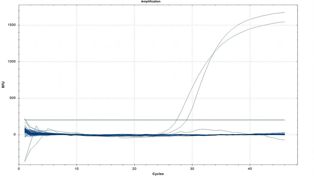
A cross-sectional study was conducted in Sulaymaniyah Governorate, Iraq, in April 2025. Water and vegetable samples were collected from both urban and rural areas. DNA extraction was performed from all samples, and E. granulosus DNA was explored using a qPCR (quantitative polymerase chain reaction) assay. Sample processing included filtering water, washing vegetables, and DNA extraction under optimized conditions.
Results
A total of 245 samples, comprising 98 (40.0%) water samples and 147 (60.0%) vegetable samples, were analyzed, with 111 (45.3%) from urban and 134 (54.7%) from rural areas. Despite the comprehensive sampling, no E. granulosus DNA was detected in any sample. All control reactions yielded positive results, but no amplification was observed in the field samples, indicating the absence of E. granulosus contamination.
Conclusion
This study found no evidence of E. granulosus DNA in water or vegetable samples from Sulaymaniyah, Iraq, suggesting a low likelihood of environmental contamination in this region. but seasonal changes, the restricted sample size, and methodological limitations mean that the presence of contamination cannot be completely excluded.
Introduction
Echinococcosis is a globally prevalent zoonotic disease caused by the larval or adult stages of tapeworms belonging to the genus Echinococcus (family Taeniidae). Recognized as one of the oldest documented human infections, its history dates to Hippocrates. The two primary forms affecting humans are cystic echinococcosis, also known as hydatid disease or hydatidosis, caused by Echinococcus granulosus, and alveolar echinococcosis, caused by Echinococcus multilocularis [1].
Echinococcosis has a worldwide distribution, with higher prevalence reported in areas such as Eastern Europe, South Africa, the Middle East, South America, Australia, and the Mediterranean, where livestock farming is widespread [1]. In some communities, the human incidence exceeds 50 cases per 100,000 person-years, with prevalence rates ranging from 5% to 10%. The World Health Organization (WHO) identifies CE as one of the major foodborne parasitic diseases globally [2].
Domestic dogs are the main definitive hosts for both Echinococcus species and represent the greatest risk for transmitting cystic and alveolar echinococcosis to humans. Dogs acquire infection by consuming livestock offal containing hydatid cysts and subsequently shed parasite eggs in their feces, which contaminate the environment, including soil, water, and grazing areas. Livestock become infected by ingesting these eggs while grazing, and humans are typically infected through the consumption of contaminated food or water [1].
Hydatid cysts can develop in nearly any organ, though the liver is most commonly affected (approximately 75%), followed by the lungs (15%), with rare involvement of the brain (2%) and spine (1%). Early stages of infection are often asymptomatic, with clinical manifestations emerging only when cysts enlarge or become complicated [3].
Environmental contamination with Echinococcus eggs can be substantial in endemic areas with high definitive host prevalence, creating a risk for human infection. However, contamination through food and the environment has long been overlooked, even though ingestion of infective eggs from these sources is the primary route of human infection [4].
This study aimed to detect the presence of E. granulosus DNA (deoxyribonucleic acid) in water and vegetable samples collected from both urban and rural areas of Sulaymaniyah Governorate, Iraq, using a sensitive qPCR (quantitative polymerase chain reaction) assay. All references were assessed using trusted predatory journal lists to verify their scholarly integrity [5].
Methods
Study design
A cross-sectional study design was employed. Water and vegetable samples were collected in April 2025 from multiple sites across Sulaymaniyah Governorate, Iraq. Sampling sites were randomly selected across the area to ensure the representativeness of both urban and rural areas. Sampling was conducted under sterile conditions, and all collected material was immediately transferred to the molecular biology department at Smart Health Tower for laboratory processing.
Sample collection
Water samples were collected directly from rivers, irrigation canals, and storage sources into sterile polypropylene containers. For each randomly selected site, one liter of water was obtained using sterile polypropylene containers. Vegetable samples, including commonly consumed leafy greens and root crops. Approximately 200 g per sample was collected using sterile gloves and placed in clean polyethylene bags. All containers and bags were clearly labeled with the date, site of collection, and sample type. Samples were maintained at 4–8 °C in insulated boxes with ice packs and processed within 6 h of collection.
Sample processing
Upon arrival at the laboratory, water samples were filtered through sterile membrane filters to concentrate parasitic elements. The residues retained on the filters were rinsed with sterile phosphate-buffered saline (PBS) (pH 7.4) and collected for downstream analysis. Vegetable samples were washed by agitation in 500 mL sterile PBS for 10 minutes at 150 rpm. The wash solutions were collected and centrifuged at 3,000 × g for 10 minutes. Resulting pellets were retained for downstream molecular examination.
To enhance DNA extraction efficiency, the pellets were subjected to mechanical disruption using a sterile mortar and pestle under liquid nitrogen, followed by three freeze–thaw cycles between liquid nitrogen (–196 °C) and a 37 °C water bath. This process facilitated rupture of eggshells or larval teguments, releasing nucleic acids for downstream extraction. The homogenate was then processed immediately for DNA extraction.
DNA extraction
Genomic DNA was extracted using the PureLink Genomic DNA Mini Kit (K1820-01, Invitrogen, Thermo Fisher Scientific, USA) according to the manufacturer’s instructions, with modifications adapted for E. granulosus. The procedure included sequential steps of lysis, binding, washing, and elusion. During the lysis step, the processed samples were incubated with Genomic Lysis/Binding Buffer and Proteinase K at 55–60 °C until complete digestion was achieved. RNase A was added during this stage to eliminate RNA contaminants. Following lysis, ethanol was added to the lysates, which were then transferred to silica-based spin columns. The washing step involved sequential treatment with PureLink Wash Buffer 1 and Wash Buffer 2 to remove salt, proteins, and other contaminants. A final dry spin was performed to eliminate residual ethanol. For elusion, PureLink Genomic Elution Buffer was added to the columns, and DNA was released into sterile collection tubes following centrifugation.
DNA quality assessment
The quality and concentration of the extracted DNA were evaluated using a spectrophotometer (NanoDrop, Thermo Fisher Scientific, USA). Absorbance was measured at 260 nm and 280 nm to determine purity. Purity assessment was performed by calculating the A260/A230 ratio, with acceptable values ranging between 1.9 and 2.2. DNA integrity was further confirmed by electrophoresis on a 1% agarose gel prepared in 1X Tris-borate-EDTA buffer and stained with ethidium bromide. Bands were visualized under ultraviolet illumination, and intact high-molecular-weight bands were taken as indicators of high-quality genomic DNA.
Preparation of primers and probes
Primers and probes were prepared according to the manufacturer’s instructions (Sigma-Aldrich). Stock solutions for each primer and probe were resuspended in nuclease-free water, briefly vortexed, and centrifuged. Working solutions were prepared by diluting the stock solutions 1:10 in nuclease-free water.
qPCR reaction mixture preparation
Luna Universal Probe qPCR Master Mix and other reaction components were thawed at room temperature and then placed on ice. Each component was briefly mixed by inversion and gentle vortexing. Reaction mixtures were prepared for the required number of samples, including 10% overage. For each 20 µL reaction, the following components were combined: 10 µL Luna® Universal Probe qPCR Master Mix (2X), 1 µL Cox3 forward primer (10 µM), 1 µL Cox3 reverse primer (10 µM), 0.5 µL Cox3 probe-FITC (10 µM), 1 µL template DNA (20–100 ng/µL), and 6.5 µL nuclease-free water (Table 1) [6,7]. The mixture was gently mixed and centrifuged briefly to collect the solution at the bottom of the tube. Aliquots were dispensed into qPCR tubes or plates, and DNA templates were added. Tubes or plates were spun briefly at 2,500–3,000 rpm to remove bubbles.
Target Species |
Target gene |
Primer and probe |
GenBank reference |
Oligonucleotide sequence (5’–3’) |
Target size (bp) |
Reference |
Echinococcus granulosus s.s |
Cytochrome oxidase subunit III |
Eg_cox3_Forward |
AF297617.1(UK) (Le et al. 2002) [7] |
TATCTGTAACACCACAAAACTCAAACC |
149 | Knapp et al. 2023 [6] |
Eg_cox3_Reverse |
CGTTGGAGATTCCGTTTGTTG |
|||||
Eg_cox3_Probe |
AACAAAAGCAAATCACAACAACGTCAACCC |
qPCR cycling conditions
Real-time qPCR was performed using the following thermal profile: initial denaturation at 95 °C for 60 seconds, followed by 40–45 cycles of 95 °C for 15 seconds and 60 °C for 30 seconds, with fluorescence readings collected at the extension step in the Fluorescein Isothiocyanate (green) channel.
Data analysis and standard curve
qPCR data were analyzed according to the instrument manufacturer's instructions. Standard curves were generated by plotting the logarithm of input DNA concentrations against Cq values to determine reaction efficiency, which was considered acceptable between 90–110% (slope –3.6 to –3.1). Correlation coefficients (R²) ≥ 0.98 were accepted. Specificity was verified by ensuring a Cq difference of ≥3 between template-containing and non-template controls. Method detection limits were determined using eight serial dilutions of the initial DNA concentration (100 ng/µL) to assess assay sensitivity. Samples were evaluated relative to standard curves and control reactions, with appropriate dilution factors accounted for.
Results
A total of 245 samples were collected and analyzed, comprising 98 (40.0%) water samples and 147 (60.0%) vegetable samples. Of these, 111 (45.3%) were obtained from urban areas and 134 (54.7%) from rural areas. Geographically, 119 (48.6%) samples were collected from the eastern region, 21 (8.6%) from the western region, 56 (22.8%) from the northern region, and 49 (20.0%) from the southern region of Sulaymaniyah Governorate (Table 2).
The qPCR assay demonstrated clear amplification in the positive controls, which produced sharp exponential fluorescence curves crossing the threshold between cycles 28 and 30. In contrast, all field samples, including both water and vegetables, exhibited flat baseline fluorescence with no detectable signal above the threshold, confirming the absence of amplification (Figure 1). Therefore, none of the 245 field samples tested positive for E. granulosus DNA.
Parameters |
Frequency (%) |
Residence |
|
Urban |
111 (45.3) |
Rural |
134 (54.7) |
Sample type |
|
Water |
98 (40.0) |
Vegetables |
147 (60.0) |
Region |
|
East |
119 (48.6) |
West |
21 (8.6) |
North |
56 (22.8) |
South |
49 (20.0) |
Discussion
Fresh vegetables are a vital part of a nutritious diet, but they may also act as carriers of protozoan cysts and helminth eggs or larvae. The moist conditions required for their growth create a favorable environment for the persistence and transmission of enteroparasitic forms. In many developing areas, the use of irrigation water contaminated with human or animal feces has been recognized as a key factor contributing to high levels of vegetable contamination with helminth eggs [8].
In this cross-sectional study of 245 water and vegetable samples collected from urban and rural areas of Sulaymaniyah, Iraq, no E. granulosus DNA was detected by qPCR. This absence of positive findings contrasts with reports from other regions documenting occasional environmental contamination. For example, Barosi and Umhang (2024) reported the presence of E. granulosus and E. multilocularis eggs in several environmental matrices, including water, soil, vegetables, and berries, with prevalence rates varying considerably [4]. Similarly, the international multicenter MEmE project detected E. granulosus sensu lato DNA in 1.3% of lettuce samples across Europe, with much higher levels reported in certain non-European countries, such as 12% of lettuce in Tunisia [9]. In that study, all positive samples were identified as E. granulosus sensu stricto, the genotype commonly associated with sheep, which confirmed that parasite eggs can adhere to food crops. The same project also recorded particularly high contamination rates in berries, with E. granulosus sensu stricto DNA found in 12% of blueberries from Pakistan and in 81.3% of strawberries from Tunisia [9]. In contrast, this study’s samples yielded no positives, suggesting that contamination of water and vegetables with E. granulosus eggs in Sulaymaniyah may be substantially lower than in the regions surveyed elsewhere.
Regional comparisons also demonstrate variability. In Iran, a neighboring country where hydatidosis is endemic, field surveys using microscopic egg detection have identified taeniid eggs (which include E. granulosus) on fresh produce at notable rates. A large Iranian study reported contamination in 7.8% of more than 2,700 vegetable samples, with lettuce showing the highest frequency [10]. In Shiraz, located in southern Iran, taeniid eggs were detected in 2.5% (2/80) of vegetables from markets and in 4.1% (6/144) of those collected directly from farms [11]. In Qazvin Province, contamination was found in 1.8% of 218 vegetable samples, again involving Taenia/Echinococcus eggs [12]. However, all these surveys relied on microscopy and did not specifically confirm E. granulosus by molecular methods; to date, no study has definitively documented E. granulosus DNA in Iranian produce despite the microscopy-based evidence [13]. In Turkey, environmental investigations have focused mainly on definitive hosts. For example, one survey of red fox feces reported 0.5% positivity for E. granulosus [14].
A few studies outside the Middle East provide context for our negative results. Awosanya et al. (2022) detected E. granulosus sensu lato DNA in environmental samples from Nigeria, with 2% of irrigation water and 7% of soil samples testing positive by PCR [15]. In Japan, Mori and colleagues analyzed river water using environmental DNA methods and detected E. multilocularis DNA in only 0.78% of samples [16], showing that such approaches often yield very low detection rates even in endemic regions. Taken together, these studies indicate that although Echinococcus eggs can contaminate water and crops, detection rates are often low unless contamination levels are substantial.
One possible explanation for the absence of positive findings in this study is the ecological and agricultural context. Environmental conditions in Sulaymaniyah, including climate, farming practices, and seasonality, may have reduced the likelihood of contamination. Experimental studies show that E. granulosus eggs can survive for more than 200 days at 7 °C under humid conditions, about 50 days at 21 °C with low humidity, and only a few hours in hot, dry conditions around 40 °C. Temperature and humidity strongly influence egg infectivity, and the eggs are highly sensitive to desiccation [17]. In Iraqi Kurdistan, late April corresponds to spring, with moderate average temperatures (~22 °C) that could support egg survival. However, rainfall or irrigation may wash eggs away, and dry days may accelerate desiccation. Agricultural practices may also play a role. For instance, if vegetables in Sulaymaniyah (such as irrigated greens) are cultivated and washed in ways that limit contact with dog feces, contamination would be less likely. In addition, the use of treated water or protected cultivation methods would further reduce exposure risk. In contrast, the high detection rates reported in countries such as Tunisia and Pakistan may reflect open-field agriculture combined with climatic or hygiene conditions more favorable to egg persistence.
This study has several important limitations. First, the sample size of 245 and the sampling strategy may have been insufficient to detect low-prevalence contamination. Although 245 samples represent a moderate number, environmental egg contamination could be so sparse that even this number yielded no positives. Second, sampling was conducted at a single time point at the end of April, so seasonal variations were not captured. E. granulosus transmission can be seasonal; for example, dog infections may peak after livestock slaughter in winter. Therefore, a late-spring snapshot may have missed periods of higher contamination or, conversely, periods when spring rains already washed eggs away.
Geographically, the study was confined to Sulaymaniyah governorate, encompassing both urban and rural areas. This region may not represent the entire area of Iraqi Kurdistan or Iraq, or neighboring provinces. As a result, the negative findings cannot be generalized to broader regions or other habitat types.
Despite these limitations, the results are reassuring from a public health perspective, suggesting that under current conditions, contaminated water and vegetables may not constitute a significant route of E. granulosus transmission in Sulaymaniyah. However, other transmission routes, particularly direct contact with infected dogs or handling of contaminated soil, remain relevant. Preventive strategies should therefore continue to focus on regular deworming of dogs, safe disposal of livestock offal, public education on hand and food hygiene, and improvements in slaughterhouse sanitation [1].
Future studies should expand sampling to cover different seasons, larger sample sizes, and additional environmental matrices such as soil and dog feces. Incorporating assays for egg viability alongside qPCR would help determine whether detected DNA reflects viable, infectious eggs or degraded material. Integrating environmental monitoring with veterinary and human epidemiological data will provide a more comprehensive understanding of local transmission dynamics, ensuring that cystic echinococcosis control efforts in Sulaymaniyah remain evidence-based and effective.
Conclusion
No E. granulosus DNA was detected in the water and vegetable samples collected from Sulaymaniyah, Iraq. These results indicate a low likelihood of environmental contamination during the study period, but seasonal changes, the restricted sample size, and methodological limitations mean that the presence of contamination cannot be completely excluded.
Declarations
Conflicts of interest: The authors have no conflicts of interest to disclose.
Ethical approval: Not applicable.
Patient consent (participation and publication): Not applicable
Source of Funding: Smart Health Tower.
Role of Funder: The funder remained independent, refraining from involvement in data collection, analysis, or result formulation, ensuring unbiased research free from external influence.
Acknowledgements: None to be declared.
Authors' contributions: RQS and MNH were major contributors to the study's conception and to the literature search for related studies. HAN, AMM, and MMA were involved in the literature review, study design, and writing of the manuscript. SJQ, AKH, SAF, SMA, SSA, YMM and KKM were involved in the literature review, the study's design, and data collection. TOM, SLE, and HSA were involved in the literature review, the study's design, the critical revision of the manuscript, and the table processing. All authors have read and approved the final version of the manuscript. RQS and HAN confirm the authenticity of all the raw data.
Use of AI: ChatGPT-4.0 was used to assist in language editing and improving the clarity of the manuscript. All content was reviewed and verified by the authors. Authors are fully responsible for the entire content of their manuscript.
Data availability statement: Not applicable.

Carcinoma ex Pleomorphic Adenoma: A Case Series and Literature Review
Abdulwahid M. Salih, Rebaz M. Ali, Ari M. Abdullah, Aras J. Qaradakhy, Ahmed H. Ahmed, Imad J....
Abstract
Introduction
Carcinoma ex pleomorphic adenoma (CXPA) is a rare malignant salivary gland tumor that can lead to severe complications and carries a risk of distant metastasis. This study aims to provide a comprehensive overview of CXPA through a case series and a review of the literature.
Methods
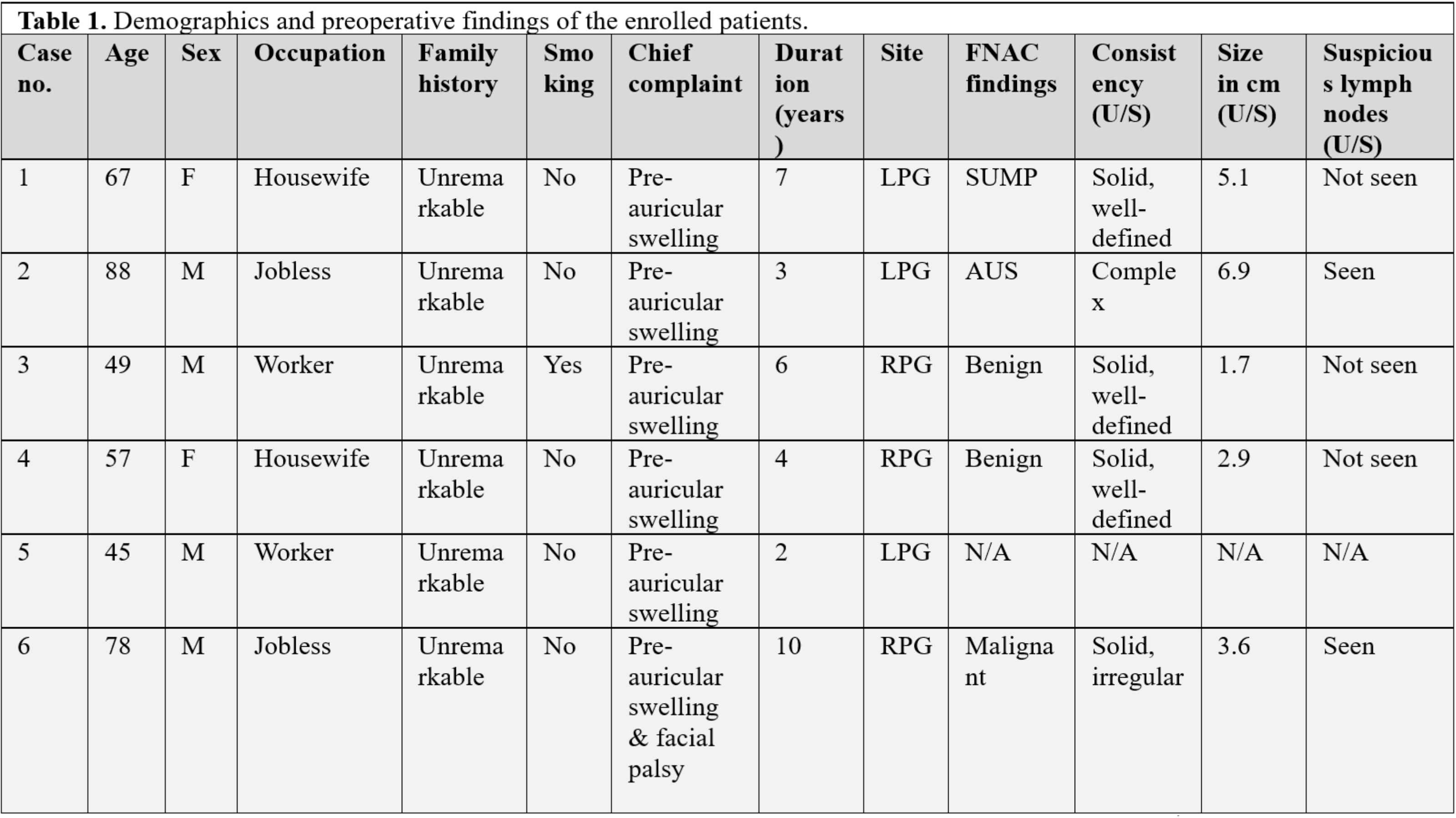
This was a single-center retrospective case series. The patients were included from November 2018 to December 2024. All confirmed cases of CXPA that were diagnosed and managed with complete clinical data were included in this study. Cases with incomplete data were excluded.
Results
Six patients were included, with ages ranging from 45 to 88 years (mean ± SD: 64 ± 15.36; median: 62). Most were male (66.7%), with an even distribution of occupations. All presented with preauricular swelling lasting 2 to 10 years, and three had left-sided tumors. Fine needle aspiration identified 33.3% as benign and 16.7% as malignant. Ultrasound examination showed solid tumors in four cases, three of which were well-defined. Three (50%) underwent total parotidectomy, and three (50%) underwent superficial parotidectomy. Histopathological examination revealed adenocarcinoma ex pleomorphic adenoma in 50% and squamous cell carcinoma ex pleomorphic adenoma in 16.7%. Tumor sizes ranged from 3.5 to 6 cm (mean: 4.73 ± 1.24 cm). Capsular invasion was present in all cases, with lymph node involvement in 33.3%, lympho-vascular invasion in 16.7%, and perineural invasion in 50%. Adjuvant therapy included radiotherapy or chemoradiotherapy.
Conclusion
Although CXPA is very rare, it is a serious condition; surgical approach with or without adjuvant therapy may result in preferable outcomes.
Introduction
Salivary gland tumors are uncommon neoplasms of the head and neck region. Among them, pleomorphic adenoma is the most prevalent benign type, accounting for approximately 70% of all salivary gland tumors [1]. The parotid gland is the most frequent site of occurrence for salivary gland tumors, followed by the submandibular gland and the minor salivary glands [2]. If left untreated, pleomorphic adenoma carries a risk of malignant transformation, with the risk reaching up to 9.5% after 15 years and continuing to increase over time [3].
Carcinoma ex pleomorphic adenoma (CXPA) is a rare malignant salivary gland tumor that develops from a pre-existing benign pleomorphic adenoma, accounting for approximately 5% of all head and neck malignancies [4]. CXPA constitutes approximately 3.6% of all salivary gland neoplasms, 6.2% of all mixed tumors, and 11.6% of all malignant salivary gland neoplasms. This malignancy predominantly occurs between the sixth and eighth decades of life and shows a slight female predominance [5]. Historical data reveal geographical variation in the incidence of this tumor relative to primary parotid malignancies, with reported rates of 12–13% in the United States, 14% in Switzerland, and up to 25% in the United Kingdom [1]. CXPA has also been referred to by other names, including carcinoma ex mixed tumor, carcinoma ex adenoma, and carcinoma ex benign pleomorphic adenoma [5].
The risk of malignant transformation is heightened by patient-related factors such as advanced age and a history of smoking, as well as disease-related factors, including larger tumor size and higher histological grade [1]. As a rare and complex disease, the clinical and pathological understanding of CXPA continues to evolve with ongoing research and advancements in diagnostic techniques [1]. This study aims to provide a comprehensive overview of CXPA by analyzing six cases, with a focus on clinical presentation, diagnostic approaches, treatment outcomes, and a review of the literature. The references have been evaluated for credibility using the most up-to-date criteria [6].
Methods
Study design
This single-center case series included consecutive patients diagnosed with CXPA who were treated between November 2018 and December 2024.
Data collection
After de-identification, the necessary data were retrospectively obtained from patient records in the Head and Neck clinic database. Extracted variables included patient demographics, occupation, clinical presentation, ultrasound (U/S) findings, treatment approach, outcomes, histopathological findings, and follow-up information. The follow-up period ranged from 1 to 5 years.
Eligibility criteria
All confirmed cases of CXPA that were diagnosed and managed with complete clinical data were included in this study. Cases with incomplete data were excluded.
Intervention
All patients underwent a thorough preoperative assessment, including a detailed clinical examination with an emphasis on facial nerve function, as well as imaging to assess lesion size, location, extent, and potential local invasion. Surgical procedures were performed under general anesthesia with the patient positioned supine and the head turned contralaterally to the lesion side. A standard Blair (lazy-S) incision was utilized, beginning anterior to the tragus, extending around the earlobe, and continuing into a natural skin crease in the upper neck to ensure optimal surgical access and cosmetic appearance.
Following subplatysmal flap elevation, dissection was carried out to identify the main trunk of the facial nerve, typically located at the stylomastoid foramen, just inferior and medial to the tympanomastoid suture. In cases of partial parotidectomy, only the superficial lobe of the gland was excised, preserving the facial nerve and its branches. In total parotidectomy, both superficial and deep lobes were removed, with caution to maintain all major branches of the facial nerve. Dissection proceeded using fine instruments and bipolar cautery under loupe magnification to enhance visualization and minimize nerve trauma.
Hemostasis was achieved using bipolar coagulation and ligation of feeding vessels. Redivac drains were placed in the surgical bed and secured with sutures to facilitate postoperative drainage and reduce the risk of hematoma or seroma formation. Skin closure was performed in layers using absorbable sutures for the deep plane and non-absorbable or subcuticular sutures for the skin to optimize healing and reduce scar formation.
Post-intervention considerations
Postoperatively, patients received protocol-based analgesia and prophylactic antibiotics. The diagnosis was confirmed through histopathological examination of the surgical specimens.
Statistical analysis
Data entry and coding were performed using Microsoft Excel 2019. Descriptive statistical analysis of qualitative data was conducted using the Statistical Package for the Social Sciences (SPSS) Version 25. Results were presented as means, frequencies, and percentages.
Results
This study included 6 patients, whose raw data are presented in Tables 1 & 2. Ages ranged from 45 to 88 years, with a median age of 62 and a mean age of 64 ± 15.36 years. Most of the patients were males (66.67%), with occupations evenly distributed (33.33% housewives, 33.33% unemployed, and 33.33% workers). Family history was unremarkable in all patients. Smoking was reported in only 1 patient. All the patients presented with preauricular swelling, with duration of symptoms ranging from 2 to 10 years. The left parotid gland was affected in 3 (50%) patients. Upon fine needle aspiration (FNA), 2 (33.33%) of the tumors were benign, while only 1 (16.67%) tumor was malignant with certainty. On U/S, 4 of the tumors were solid, 3 of which were well-defined. Lymph nodes were suspicious for involvement in 2 (33.33%) patients. three patients (50%) underwent total parotidectomy, while the other 3 (50%) underwent superficial parotidectomy. Histopathology revealed adenocarcinoma ex pleomorphic adenoma in 3 (50%) patients, and squamous cell carcinoma ex pleomorphic adenoma in 1 (16.67%) patient. Tumor sizes ranged from 3.5 to 6 cm (mean: 4.73 ± 1.24). Lymph node involvement was seen in 2 (33.33%) patients. Capsular invasion was observed in all patients, lympho-vascular invasion in 1 (16.67%) patient, and perineural invasion in 3 (50%) patients. Adjuvant therapy included radiotherapy and chemoradiotherapy (Table 3). No cases of recurrence were reported, and one patient passed away from old age.
Case no. |
Age |
Sex |
Occupation |
Family history |
Smoking |
Chief complaint |
Duration (years) |
Site |
FNAC findings |
Consistency (U/S) |
Size in cm (U/S)
|
Suspecious lymph nodes (U/S) |
1 |
67 |
F |
Housewife |
Unremarkable |
No |
Pre-auricular swelling |
7 |
LPG |
SUMP |
Solid, well-defined |
5.1
|
Not seen |
2 |
88 |
M |
Jobless |
Unremarkable |
No |
Pre-auricular swelling |
3 |
LPG |
AUS |
Complex |
6.9
|
Seen |
3 |
49 |
M |
Worker |
Unremarkable |
Yes |
Pre-auricular swelling |
6 |
RPG |
Benign |
Solid, well-defined |
1.7
|
Not seen |
4 |
57 |
F |
Housewife |
Unremarkable |
No |
Pre-auricular swelling |
4 |
RPG |
Benign |
Solid, well-defined |
2.9
|
Not seen |
5 |
45 |
M |
Worker |
Unremarkable |
No |
Pre-auricular swelling |
2 |
LPG |
N/A |
N/A |
N/A
|
N/A |
6 |
78 |
M |
Jobless |
Unremarkable |
No |
Pre-auricular swelling & facial palsy |
10 |
RPG |
Malignant |
Solid, irregular |
3.6 |
Seen |
F: Female, M: Male, LPG: Left parotid gland, RPG: Right parotid gland, SUMP: Salivary gland Neoplasm of Uncertain Malignant Potential, AUS: Atypia of Undetermined Significance, U/S: Ultrasonography |
Surgical approach |
HPE |
Tumor stage |
Tumor size (CM) |
LN involvement |
Invasion |
Adjuvant therapy |
Outcome |
Follow-up (years) |
|||
Capsular |
Lympho-vascular |
Perineural |
|||||||||
Total Parotidectomy |
Adenocarcinoma ex pleomorphic adenoma |
pT3 |
5.5 |
N/A |
FI |
Not seen |
Not seen |
N/A |
No recurrence |
4 |
|
Superficial parotidectomy, suprahyoid lymph nodes dissection & excision of sublingual gland |
Adenocarcinoma ex pleomorphic adenoma |
|
6 |
Seen |
I |
Not seen |
Seen |
Radiotherapy |
Died |
N/A |
|
Superficial parotidectomy |
Squamous cell carcinoma ex pleomorphic adenoma |
pT2 N0 |
3.5 |
Not seen |
I |
Not seen |
Not seen |
N/A |
No recurrence |
5 |
|
Superficial parotidectomy |
Carcinoma ex-pleomorphic adenoma |
pT2 N0 R0 |
3.5 |
Not seen |
FI |
Not seen |
Not seen |
CCRT |
No recurrence |
2 |
|
Left total parotidectomy |
Carcinoma ex-pleomorphic adenoma |
pT2 N0 |
3.9 |
Not seen |
FI |
Not seen |
Seen |
CCRT |
No recurrence |
1 |
|
Right total parotidectomy with right cervical lymph node dissection |
Adenocarcinoma ex pleomorphic adenoma |
pT4a N3b R1 |
6 |
Seen |
I |
Extensive |
Seen |
CCRT |
No recurrence |
1 |
Variables |
Frequency/percentage |
Sex Male Female |
Total (6) 4 (66.67%) 2 (33.33%) |
Age (years) Range Mean (±SD) Median (IQR) |
45-88 64 ± 15.36 62 (29) |
Smoker Non-smoker |
1 (16.67%) 5 (83.33%) |
Chief complaint Preauricular swelling Preauricular swelling & facial palsy |
5 (83.33%) 1 (16.67%) |
Duration of symptoms (years) Range Mean (±SD) |
2-10 5.3 ± 2.69 |
Site Left parotid gland Right parotid gland |
3 (50%) 3 (50%) |
Surgical approach Total parotidectomy Superficial parotidectomy |
3 (50%) 3 (50%) |
Adjuvant therapy Radiotherapy Concurrent chemo-radiotherapy N/A |
1 (16.67%) 3 (50%) 2 (33.33%) |
HPE Adenocarcinoma ex pleomorphic adenoma Squamous cell carcinoma ex pleomorphic adenoma Carcinoma ex pleomorphic adenoma |
3 (50%) 1 (16.67%) 2 (33.33%) |
Degree of invasion Low degree Medium degree High degree |
3 (50%) 1 (16.67%) 2 (33.33%) |
Invasion status Capsular Lympho-vascular Perineural |
6/6 (100%) 1/6 (16.67%) 3/6 (50%) |
Tumor size (cm) Range Mean (±SD) |
3.5-6 4.73 ± 1.24 |
Outcome No recurrence Death |
5 (83.33%) 1 (16.67%) |
SD: Standard deviation, IQR: Interquartile range, N/A: Not available, HPE: Histopathology |
Discussion
The median age of this study was 62 years, which is close to a study by Suzuki et al., which had a median age of 60 years [7]. The mean age of 64 in the current study was also comparable to other studies. For example, a retrospective study of 73 patients with CXPA had a mean age of 61 years [8]. However, a lower mean of 55.1 was reported by Seok et al. [9]. Kato et al. reported four cases, one of which involved a 48-year-old individual [3]. Recently, another case of CXPA in a 45-year-old male was also reported [10]. When compared to pleomorphic adenoma, the mean age of diagnosis is shown to be higher by 13 years [9].
The current series demonstrated a male predominance, consistent with findings from a systematic review by Key et al., in which males accounted for 58.9% of cases [1]. In this series, the parotid gland was involved in 100% of cases, higher than the typical range reported in the literature, which may reflect referral bias. Nevertheless, the parotid remains the most commonly affected salivary gland subsite [1].
Patients presented with symptom durations ranging from 2 to 10 years, which is notably shorter than what is commonly reported in earlier literature. A comprehensive review, for example, documented a mean symptom duration of 23.3 years [5]. Longer durations reaching 50 years have also been reported [3]. However, more recent reports indicate shorter intervals; Keerthi et al. reported a case with only six months of symptoms [11]. The short symptom durations in the current study may reflect earlier detection and referral patterns or more aggressive tumor biology, leading to earlier presentation.
One of the diagnostic challenges was the low sensitivity of FNA for malignancy at 16.67%, which is a known limitation of FNA in CXPA diagnosis. In a series of 16 patients, only seven (43%) were identified as malignant [12]. In a cohort of 260 patients, 170 patients (65.4%) had a preoperative FNA. In 156 of those (91.8%), the FNA diagnosed a benign tumor, with the rest having an unsatisfactory or nondiagnostic FNA [13]. Parotid FNA carries two potential sources of false-negative results: sampling a benign area of a pleomorphic adenoma rather than the malignant component, and misclassifying a low-grade CXPA as a benign pleomorphic adenoma [13].
Tumor sizes in reported cases range from 1 to 26 cm [4]. Similarly, the sizes observed in the present study fall within this typical range. A multivariate analysis has identified tumor size ≥4 cm as a factor associated with worse prognosis. Notably, six patients in the current series (66.7%) had tumors larger than 4 cm but demonstrated favorable outcomes, suggesting that effective treatment protocols may mitigate the impact of tumor size on prognosis.
Recent studies have shown that the most frequently encountered histological types in CXPA are highly malignant adenocarcinoma and undifferentiated carcinoma. However, a wide spectrum of other subtypes has also been reported, including squamous cell carcinoma, mucoepidermoid carcinoma, adenoid cystic carcinoma, papillary carcinoma, and terminal duct carcinoma [11]. In the current series, adenocarcinoma accounted for 50% of cases, while squamous cell CXPA was observed in one patient. This aligns with findings from a retrospective study of 24 patients, which reported six cases of adenocarcinoma and a single case of squamous cell carcinoma [12].
Certain pathological features, such as invasiveness, have been studied in the literature. In particular, extracapsular extension and invasion measuring ≥1.5 mm have been linked to an increased risk of recurrence and mortality [1]. Capsular invasion was present in all cases in the current series, with 50% showing focal infiltration. Lympho-vascular invasion was observed in 16.67%, and perineural invasion in 50%. Similar findings have been reported in the literature, such as Kim et al.'s study of 17 CXPA patients, where lympho-vascular invasion was seen in three patients [14]. In a cohort of 215 patients with parotid gland tumors, 14 of whom were diagnosed with CXPA, perineural invasion was documented in 21.4% [15]. In a retrospective study of 51 patients, 45.1% exhibited perineural invasion [16]. Another study involving 37 CXPA patients found 43% with perineural invasion and 40.5% with lympho-vascular invasion [17]. Perineural invasion significantly impacts distant metastasis, tumor-specific survival, and overall survival (P < 0.05), and tends to be associated with locoregional recurrence (P = 0.086). In multivariate analysis, perineural invasion is identified as an independent prognostic factor for overall survival [16].
Lymph node invasion was observed in 33.3% of the patients, which is comparable to findings from previous studies. For instance, a review involving 619 patients reported lymph node involvement, both single and multiple nodes, in 29.6% of cases [18]. Another retrospective analysis of 51 patients revealed that 33.3% of the patients had lymph node involvement [16]. In their study, Zhao et al. identified advanced T stage and lymph node involvement as important factors for an unfavorable clinical outcome [16].
Treatment of CXPA involves an ablative surgical procedure, which may or may not be followed by reconstructive surgery [5]. To date, no universally accepted treatment protocol exists for this tumor type [4]. Zhao et al. emphasized that the extent of surgical intervention should be individualized, taking into consideration the tumor’s location, size, and the involvement of adjacent anatomical structures. For parotid gland tumors, a total or radical parotidectomy is generally recommended for frankly invasive CXPA, with facial nerve resection indicated if direct tumor infiltration is evident [16]. In the present series, three patients underwent superficial parotidectomy, one of whom also underwent suprahyoid lymph node dissection and excision of the sublingual gland due to regional extension, and the other three underwent total parotidectomy.
Adjuvant therapies for CXPA may include radiotherapy or chemotherapy, primarily aimed at improving local control and potentially enhancing survival outcomes [10]. However, due to the rarity of the disease, data on the specific efficacy of radiotherapy are limited [16]. In a retrospective analysis of 63 patients, Chen et al. reported that postoperative radiotherapy significantly improved local disease control, although it did not confer a clear survival benefit [19]. Chemotherapy is generally reserved for patients with advanced, recurrent, or metastatic disease and is used primarily for palliative purposes. The role of radiotherapy remains controversial and is typically considered in cases with high-grade histology, positive margins, perineural invasion, or lymph node involvement [20]. Historically, CXPA has been regarded as a high-grade malignancy, often necessitating adjuvant radiotherapy. This classification is reflected in data from a national American cancer database, which demonstrated that a higher proportion of patients with CXPA were selected to receive chemoradiotherapy [1]. Three patients received adjuvant chemoradiotherapy, all of whom had good outcomes.
Patients diagnosed with noninvasive or minimally invasive CXPA generally exhibit favorable outcomes, with low recurrence rates and minimal risk of metastasis. In contrast, those with frankly invasive tumors have a significantly poorer prognosis. Reported recurrence rates for invasive CXPA range from 23% to 50%, and distant metastases may occur in up to 70% of cases, reflecting the aggressive biological behavior of the invasive subtype [4]. No recurrences were observed in this series. While this may indicate effective treatment approaches, it could also be due to the relatively short follow-up periods.
Conclusion
Although CXPAs are very rare, they are serious conditions that may originate from benign conditions. Surgical approach with or without adjuvant therapy might result in good outcomes.
Declarations
Conflicts of interest: The authors have no conflicts of interest to disclose.
Ethical approval: The study was ethically reviewed and approved by the Scientific Committee of the Kscien organization (Approval No. 2025-38).
Patient consent (participation and publication): Not applicable
Source of Funding: Smart Health Tower.
Role of Funder: The funder remained independent, refraining from involvement in data collection, analysis, or result formulation, ensuring unbiased research free from external influence.
Acknowledgements: None to be declared.
Authors' contributions: AMS and SHH were major contributors to the study's conception and to the literature search for related studies. AMA was the pathologist who performed the histopathological diagnosis. AJQ and AHA were the radiologists who performed and assessed the cases. MMA, HAA, and AAQ were involved in the literature review, study design, and writing of the manuscript. RMA, IJH, STSA, and MLF were involved in the literature review, the study's design, the critical revision of the manuscript, and the table processing. AMS and MMA confirm the authenticity of all the raw data. All authors have read and approved the final version of the manuscript
Use of AI: ChatGPT-4.0 was used to assist in language editing and improving the clarity of the manuscript. All content was reviewed and verified by the authors. Authors are fully responsible for the entire content of their manuscript.
Data availability statement: Not applicable.

Perceptions of Telemedicine and Rural Healthcare Access in a Developing Country: A Case Study of Bayelsa State, Nigeria
Ebidor Lawani-Luwaji, Chibuike Frederick Okafor
Abstract
Introduction
Telemedicine is the remote delivery of healthcare services using information and communication technologies and has gained global recognition as a solution to address healthcare disparities. This study explores the perceptions of telemedicine and its potential to improve rural healthcare access in a developing country through the insights of undergraduate Medical Laboratory Science students.
Methods
A descriptive cross-sectional study was conducted among 42 fourth-year students of the Medical Laboratory Science program. Respondents completed a structured questionnaire that assessed their awareness, familiarity, perceived benefits and barriers to telemedicine, and their views on its applicability in rural Bayelsa.
Results
The findings indicated that while the majority of respondents (60.5%) were aware of telemedicine, their understanding of specific types, such as asynchronous and synchronous telehealth, was limited. The main perceived benefits were improved healthcare access (48.8%) and reduced costs (18.6%). Acceptance levels varied, with 47.6% endorsing telemedicine, while others remained uncertain or sceptical.
Conclusion
The study reveals enthusiasm and knowledge gaps among future healthcare professionals regarding telemedicine. It highlights the need for targeted education, digital literacy, and infrastructure investment to enable telemedicine in rural Nigerian communities.
Introduction
Telemedicine, the remote delivery of healthcare services using information and communication technologies (ICTs), has emerged as a transformative strategy for improving healthcare access, particularly in rural and underserved regions [1]. Overcoming geographical and logistical barriers enables timely consultations, diagnostics, and follow-up care without requiring long-distance travel.
In many developing countries, telemedicine has become an increasingly important solution for bridging healthcare gaps caused by workforce shortages, underdeveloped infrastructure, and rural-urban healthcare disparities. Low- and middle-income countries (LMICs) in Africa and Asia have seen gradual adoption of digital health technologies, including mobile health (mHealth) platforms and virtual consultations, especially during and after the COVID-19 pandemic [2]. While countries such as Rwanda, Ghana, and India have launched national digital health strategies, challenges like unreliable internet, funding limitations, and limited technical literacy continue to affect large-scale implementation [3]. Within this global context, Nigeria shares many of these challenges but also presents unique opportunities for leveraging telemedicine in improving rural healthcare access.
In Nigeria, where disparities in healthcare access are especially pronounced in rural areas, telemedicine holds considerable promise. Previous studies have demonstrated its benefits like reduced patient travel time and improved early diagnosis rates. Other studies found that telemedicine enhanced antenatal care coverage in rural communities in Enugu. These findings emphasise the potential of telemedicine in addressing healthcare disparities in diverse Nigerian settings [4,5].
However, the country's healthcare infrastructure and human resources remain critically inadequate, with a disproportionate concentration in urban areas [6]. Rural residents, who make up over 40% of the Nigerian population, face poor access to quality and affordable healthcare. Physical distance, limited transportation, and high travel costs often delay or prevent illness treatment, especially in geographically isolated areas [7]. These challenges have resulted in adverse health outcomes, including higher rates of infectious diseases and maternal and child mortality in rural communities. Telemedicine offers an opportunity to address these inequities by connecting healthcare providers with rural populations through virtual consultations and mobile health technologies [8]. Mobile platforms, in particular, facilitate affordable and convenient care delivery in settings lacking comprehensive health infrastructure.
While telemedicine has demonstrated significant potential in rural regions across Africa, successful implementation remains context-dependent, requiring adaptation to local infrastructure and socio-cultural realities [9]. Despite its benefits, the widespread adoption of telemedicine in Nigeria remains limited due to persistent challenges such as unreliable internet connectivity, the high cost of digital tools, and low levels of digital literacy among both healthcare workers and patients [10]. These challenges are even more pronounced in Bayelsa State, a region in Nigeria's Niger Delta known for its swampy terrain, scattered riverine communities, and poor road infrastructure [11,12]. Given these constraints, Bayelsa stands out as a strong candidate for targeted telemedicine interventions. However, there is a notable gap in data regarding local perceptions and readiness, particularly in these uniquely challenging settings.
In this context, telemedicine may enhance healthcare delivery by mitigating travel-related barriers through remote diagnostics, video consultations, and mobile health applications. However, developing effective implementation strategies requires an understanding of local awareness, perceptions, and barriers to the use of telemedicine.
In Nigeria, understanding local perceptions is critical to designing effective, context-specific telemedicine interventions. This study focuses on Bayelsa State, located in the country's Niger Delta region. It explores the perceived impact of telemedicine on healthcare access through the insights of 400-level Medical Laboratory Science students at Niger Delta University. These students represent a well-educated segment of the rural population, many of whom live in or maintain close ties with underserved communities. Their clinical training equips them with a foundational understanding of healthcare systems and digital innovations, such as telemedicine, allowing them to assess its applicability critically.
Furthermore, as future healthcare professionals, their perspectives offer valuable insight into both community needs and the likelihood of professional adoption of telemedicine. Their dual role as rural community members and emerging practitioners provides a unique vantage point for evaluating the feasibility, acceptance, and scalability of telemedicine. The findings aim to inform evidence-based policy and program strategies that could enhance healthcare access in geographically disadvantaged regions like Bayelsa State.
Methods
Study area
This study was conducted among 400-level students in the Department of Medical Laboratory Science at Niger Delta University, located in Bayelsa State, Nigeria. The university is situated in a region with significant rural and riverine communities, making it relevant for healthcare access and telemedicine research.
Study design
A descriptive cross-sectional study assessed perceptions of telemedicine and its potential impact on healthcare access in rural communities.
Population and sample size
The target population comprised all 400-level students enrolled in the Department of Medical Laboratory Science at Niger Delta University. Participants were selected based on their willingness and availability to respond to the questionnaire. This population was chosen because it represents a group of emerging healthcare professionals who are academically exposed to modern healthcare systems and likely to engage with digital health solutions in their future careers. As senior students in a clinical field, their perspectives offer valuable insights into the readiness and acceptance of telemedicine among the next generation of practitioners. Furthermore, their residence in Bayelsa State ensures contextual relevance, as they can relate professionally and personally to the healthcare access challenges faced in the region.
Data collection instruments
The primary instrument for data collection was a structured questionnaire developed using Google Forms. The questionnaire consisted of both closed-ended and open-ended questions designed to assess respondents' demographics, awareness of telemedicine, perceived benefits, challenges, and preferred access modes.
Data collection procedure
The Google Form link containing the questionnaire was distributed electronically via WhatsApp to prospective participants. Respondents accessed and completed the form using their electronic devices.
Statistical analysis
Descriptive statistics were used to summarise the data. The results are presented in tables and charts to illustrate key findings clearly.
Results
The majority of respondents were aged 21–25 years, indicating a predominance of young adults (Table 1). Most respondents were female (64.3%), while males comprised 35.7%.
Age Group (Years) |
Number of Respondents |
Percentage (%) |
16–20 |
4 |
9.52% |
21–25 |
28 |
66.67% |
26–30 |
5 |
11.90% |
31–35 |
5 |
11.90% |
Total |
42 |
100% |
The most frequently reported challenge to accessing healthcare was the high cost of healthcare services, cited by 50% of respondents. Other notable barriers included a shortage of medical personnel (21.4%) and long travel distances to healthcare facilities (14.3%). A smaller proportion of respondents either did not know where to access services (2.4%) or identified other issues, such as long wait times or inadequate infrastructure (11.9%) (Table 2). When asked what came to mind when thinking of telemedicine, 60.5% selected the combination of video calls, phone calls, and health applications. Smaller groups identified health apps alone (11.6%), phone calls (4.7%), or video calls (2.3%), while 20.9% indicated they were unfamiliar with the concept (Figure 1).
Challenge |
Number of Respondents |
Percentage (%) |
Too far |
6 |
14.29% |
Too expensive |
21 |
50.00% |
No Medical personnel |
9 |
21.43% |
Do not know where to go |
1 |
2.38% |
Others |
5 |
11.91% |
Total |
42 |
100% |
In terms of familiarity with specific types of telemedicine, 58.1% of respondents reported no awareness. Among the remaining respondents, 27.9% were familiar with mobile health (mHealth), 7% with remote monitoring, 4.7% with synchronous telehealth, and 2.3% with asynchronous telehealth (Figure 2).
Regarding the perceived benefits of telemedicine, 48.8% of respondents identified increased healthcare access as an important advantage. Other selected benefits included convenience (20.9%), reduced travel time and cost (18.6%), and improved health outcomes (9.3%). Only 2.4% selected "all the options" (Figure 3).
Discussion
Nigeria's healthcare system has consistently struggled to meet the demands of its rapidly growing population, which has surpassed 200 million [13,14].
Findings revealed that most respondents recognised telemedicine as involving video calls, phone calls, and health applications, indicating a broad level of general awareness. However, knowledge of specific categories such as asynchronous and synchronous telehealth was limited. This gap between general familiarity and specific understanding suggests the need for targeted education and capacity-building to support effective adoption of telemedicine services in rural communities.
The distribution of respondents across urban and rural backgrounds further underscored the relevance of telemedicine in both settings. However, its utility is particularly critical in rural areas where persistent barriers, such as travel distance, lack of transport, and shortage of healthcare professionals, continue to hinder timely access to care [15]. These issues are especially prevalent in geographically complex regions like Bayelsa State.
Healthcare-seeking behaviour among respondents also reflected patterns consistent with young adult populations, with many reporting infrequent visits to health facilities. This may be due to a combination of factors, including cost, convenience, and perceived need. Telemedicine presents an opportunity to address this behavioural trend by offering more accessible, flexible, and digitally enabled healthcare services suitable for this demographic.
Respondents identified high healthcare costs, shortage of medical personnel, and travel distance as the main barriers to accessing care. These findings align with previous research highlighting cost and provider shortages as critical obstacles to healthcare access in Nigeria [13-16]. In Bayelsa, the challenges are compounded by infrastructural limitations and rugged terrain. Telemedicine can mitigate these access issues by facilitating remote consultations, reducing the need for travel, and expanding the reach of healthcare professionals [17].
Although nearly half of the respondents expressed positive attitudes toward telemedicine, a significant portion remained either unsure or not in support. This suggests that awareness alone does not guarantee acceptance. Other factors, such as trust in virtual consultations, digital literacy levels, and user experience, may likely influence attitudes toward adoption. Similar findings in the literature indicate that successful implementation requires more than infrastructure; it must also involve building community trust and providing user-friendly, culturally appropriate solutions [18].
Supporting evidence confirms that a deeper understanding of telemedicine correlates with greater satisfaction and willingness to use digital health services [19]. The challenges reported in this study are consistent with known access barriers, reinforcing telemedicine's potential to address the structural limitations affecting rural healthcare delivery [16,20]. Prior studies have shown that telemedicine can reduce travel time, enable early diagnoses, and increase the efficiency of healthcare systems [15,19]. These benefits are particularly valuable in low-resource settings.
Interestingly, while much of the literature highlights internet connectivity and the cost of digital tools as primary barriers, respondents in this study placed more emphasis on the affordability and availability of healthcare services themselves [9]. This suggests that for students and communities in Bayelsa, immediate access to care is a more pressing concern than the infrastructure required to support digital health. In regions where core health services are still lacking, addressing foundational healthcare needs may take precedence over technological investments.
As with all self-reported data, there is a potential for response bias influenced by individual beliefs or differing levels of exposure to digital health tools.
Conclusion
The findings of this study broadly support existing literature affirming telemedicine's potential to improve healthcare access in Nigeria. The study underscores the need for context-specific, targeted strategies in implementing telemedicine, especially approaches that address affordability, community trust, and digital literacy. Future studies should examine broader stakeholder readiness, infrastructure challenges, and user training needs to guide effective policy and implementation.
Declarations
Conflicts of interest: The authors have no conflicts of interest to disclose.
Ethical approval: This study was granted by the Research Ethics Committee of Niger Delta University.
Patient consent (participation and publication): Was obtained from all participants prior to completing the questionnaire, and participants were assured of confidentiality and anonymity.
Funding: The present study received no financial support.
Acknowledgements: None to be declared.
Authors' contributions: CFO conceptualised the idea and collected the data, while ELL wrote the manuscript and analysed the data. All authors read and approved the manuscript.
Use of AI: Grammarly was used to assist in language editing and improving clarity. All content was reviewed and verified by the authors.
Data availability statement: Not applicable.
Review Articles

Hydatid Disease of The Brain Parenchyma: A Systematic Review
Fattah H. Fattah, Azad Star Hattam, Zana Omar kak Abdullah, Khanda A. Anwar, Rezhen J. Rashid,...
Abstarct
Introduction
Isolated brain hydatid disease (BHD) is an extremely rare form of echinococcosis. A prompt and timely diagnosis is a crucial step in disease management. This study is a systematic review of studies on intra-parenchymal BHD.
Methods
Studies that had the following properties were included: 1) The intra-parenchymal brain infection had been confirmed by diagnostic modalities, surgical findings, or histopathology. 2) The patient details were provided in the study. 3) The cystic lesion [s] were located intracranially.
Results
Altogether, 112 studies with a sample size of 178 cases met the inclusion criteria. Males (60.1%) showed a higher prevalence of the disease than females (38.2%). Most of the cases (64%) were affected during the first and second decades of their lives. Left-side multi-lobe involvement was the most common type of involvement (28.1%), followed by right-side multi-lobe involvement (26.4%). Surgery was the primary treatment option (97.2%), with the Dowling technique or the modified Arana-Iniguez method as the preferred approach. The total recurrence and mortality rates were 7.3% and 3.4%, respectively.
Conclusion
The definitive treatment for BHD is surgery, with the aim of removing cysts intact or excising mass lesions completely. A history of cyst rupture during operation may increase the likelihood of recurrence, and an extensive follow-up is required.
Introduction
Hydatid disease (HD) is a parasitic infection caused by the larvae of the tapeworm Echinococcus. Different genera of this microorganism can cause disease; however, in humans, two species have major clinical sequelae. Echinococcus granulosus results in cystic disease, the most common type, while Echinococcus multilocularis causes alveolar echinococcosis (AE), presenting as a mass or cystic lesion. The latter form of the disease is more invasive and aggressive, accompanied by numerous diagnostic and management challenges [1-3]. The most common organs affected by hydatidosis are the liver and lungs. However, other parts of the body can also be affected, including the bones, pericardium, orbits, ovaries, central nervous system (CNS), and other organs. In the literature, 2–3% of cases show involvement of the CNS. The incidence of isolated brain involvement is reported to be 1–2% of all cases of echinococcosis, representing approximately 2% of all intracranial space-occupying lesions [4-6]. Brain hydatid disease (BHD) is endemic in many regions where livestock raising is prevalent, and human-animal contact is common. The incidence varies geographically, with higher rates reported in rural areas. However, globalization and increased travel have led to sporadic cases being reported in non-endemic regions as well. Humans can become infected through the ingestion of parasite eggs in contaminated food, water, or by direct contact with infected dogs, canines, and sheep [7,8]. Most cases of intracerebral echinococcosis are diagnosed in pediatrics (50-75%) [9]. The clinical presentation of hydatidosis depends on the patient's age, the size, number, and location of the cyst, as well as the host's immune system. Patients with HD can remain asymptomatic for long periods, as the lesions take years to develop. When they grow well, intracranial hypertension secondary to the mass effect on the surrounding tissues is usually the first clinical sign of brain involvement. The disease may not cause focal neurological signs until they become enlarged [10-12]. In the literature, several reviews have been published on cerebral HD; however, there is a scarcity of systematic reviews on the topic. This study is a systematic review of studies on intra-parenchymal BHD published over the last two decades [1-112].
Methods
Study design and reporting standards
The study followed the Preferred Reporting Items for Systematic Reviews and Meta-Analyses (PRISMA) guidelines.
Search strategy
A systematic review of all published studies on brain parenchymal HD was conducted from 2000 to 2024 using the following databases: Google Scholar, PubMed/MEDLINE, Cochrane Library, Science Direct, and EMBASE. The keywords used in the search included:
[brain OR intraparenchymal OR cerebral OR intracerebral OR cerebrum] AND [hydatid OR hydatidosis OR echinococcoses OR echinococcosis OR echinococcal OR echinococcus].
Eligibility criteria
Non-English language studies and those unrelated to humans were excluded before or during the initial screening. Studies of BHD were included if: 1) Diagnostic modalities, surgical findings, or histopathology confirmed the intraparenchymal brain infection. 2) Patient details were provided in the study. 3) Studies published in predatory journals (inappropriately peer-reviewed) and those not meeting inclusion criteria were excluded [113].
Study selection
Titles and abstracts of identified studies were initially screened, followed by full-text screening to assess eligibility.
Data extraction
Data extracted from eligible studies included study design, country of study, patient age, gender, residency, symptoms, medical history of HD, cyst characteristics, diagnosis, management, follow-up, and outcomes.
Data analysis
Data were analyzed qualitatively (descriptive analysis) using the Statistical Package for the Social Sciences (SPSS) version 27.0 software
Results
In total, 318 studies were obtained from the resources. Before any screening, 38 of them were directly excluded due to duplication, non-English language, non-articles, and animal studies. Following the initial screening, 92 studies did not meet the inclusion criteria and were excluded. The remaining 188 studies underwent full-text screening, and 122 of them were assessed for eligibility. Ultimately, 112 studies (comprising 178 cases) met the inclusion criteria (Figure 1). The characteristics of the included studies are shown in Table 1. Out of these studies, 101 (90.2%) were case reports, 10 (8.9%) were case series, and one (0.9%) was a retrospective cohort study. Most of the cases were reported in Turkey (24.1%), followed by Iran (16.7%), India (15.2%), and Morocco (9.8%). Males (60.1%) showed a higher prevalence of the disease than females (38.2%). Most of the cases (64%) occurred in the first and second decades of life, with a mean age of 20.44 ± 16.76 years. There were 71 cases (39. 9%) in rural areas and eight cases (4.5%) in urban areas. The residency of the remaining 99 cases (55.6%) was not reported. The type of the disease was cystic in 158 cases (88.8%) and alveolar in 20 cases (11.2%). Thirteen (7.3%) cases had a previous history of HD. The most commonly presented symptoms were signs of raised intracranial pressure, including headache (62.9%), vomiting (43.3%), followed by seizure (30.3%) and paresis (28.7%). Multiple organ involvement was present in 48 (27%) cases, involving the lung, liver, kidney, adrenal gland, blood vessels, or bones. The disease was primary with a single cyst or lesion in 118 patients (66.3%), primary with multiple cysts in 27 (15.1%), secondary with a single cyst in 23 (13%), and secondary with multiple cysts in 10 (5.6%). Left-side multi-lobe involvement was the most common type of involvement (28.1%), followed by right-side multi-lobe involvement (926.4%) and parietal lobe involvement (18.5%).
Serology had been done in 55 cases (30.9%), and it was positive in 34 (19.1%). Computed tomography scans (CT) or magnetic resonance imaging (MRI) were used in all cases. Surgery was the main treatment option (97.2%). The Dowling technique, or modified Arana-Iniguez, was the method of choice (95.5%). Surgery in three cases (1.7%) was done through the Burr-hole technique instead of open craniotomy. Conservative management was performed in five cases (2.8%). The patients underwent follow-up with a mean interval of one year. Recurrence was reported in 13 cases (7.3%). Among those, six cases (46.1 %) had intra-operative complications of traumatic rupture of the cyst, and two cases (15.4 %) had a surgical puncture of the cyst. The remaining five cases (38.5%) did not experience any intraoperative complications. The mortality rate was 3.4% (Table 2).
Author |
Country |
Study design |
No |
Age |
Sex |
Presenting symptoms |
Imaging |
ISHC |
No. of cyst [s] in brain |
Location of cyst [s] in brain |
Size [cm] |
Serology |
Type of management |
Pre-Op complication |
Intra-Op complication |
Post-Op complication |
Adjuvant therapy |
Follow up* outcome |
Svrckova et al [1] |
United Kingdom
|
Case report
|
3 |
30 |
M |
Headache, seizure |
MRI |
Yes |
>1 |
Right parietal, right temporal |
N/A |
Positive |
Conservative [Albendazole/praziquantel/steroid/antiepileptic] |
N/A |
N/A |
N/A |
None |
Improved |
26 |
M |
Collapse, slurred speech, seizure, left side hemiparesis |
CT, MRI |
Yes |
1 |
Right parietal and basal ganglia |
N/A |
Positive |
Conservative [Albendazole/Praziquantel/steroid/Antiepileptic] |
N/A |
N/A |
N/A |
None |
Improved |
||||
37 |
M |
Dry cough |
MRI |
Yes |
>1 |
Bilateral hemisphere |
N/A |
Positive |
Conservative [Albendazole] |
N/A |
N/A |
N/A |
None |
Improved |
||||
Altibi et al [2] |
Brazil |
Case report |
1 |
13 |
M |
Headache, nausea |
CT, MRI |
Yes |
1 |
Right parieto-occipital |
4.7 |
Negative |
Surgical removal [Dowling]/neuronavigation |
None |
None |
None |
N/A |
N/A |
Casulli et al [3] |
Italy |
Case report |
1
|
6 |
M |
Right side hemiparesis |
CT, MRI |
Yes |
1 |
Left fronto-parietal |
6.8 |
Negative |
Surgical removal/neuronavigation |
None |
None |
Seizure, headache, worsened right hemiparesis, peri-lesional edema |
Albendazole, Antiepileptic,Steroid |
Improved |
Lakhdar et al [4] |
Morocco |
Case report |
1 |
30 |
M |
Headache, right side hemiparesis |
MRI |
Yes |
>1 |
Left fronto-parietal |
N/A |
Negative |
Surgical removal |
None |
Rupture of cysts |
None |
Albendazole, Antibiotics, Antiepileptic |
Recovered |
Fariba Bi. [5] |
Iran |
Case report |
1 |
18 |
F |
Headache, nausea, vomiting |
MRI |
Yes |
1 |
Right temporal |
N/A |
N/A |
Surgical removal |
None |
None |
None |
Albendazole, anticonvulsant |
Recovered |
Saleh et al [6] |
Egypt |
Case series
|
4 |
9 |
M |
Drowsiness, vomiting, blurred vision, headache |
CT, MRI |
Yes |
>1 |
Right parieto-occipital |
N/A |
N/A |
Surgical removal [Dowling] |
None |
None |
None |
Albendazole |
N/A |
10 |
M |
Seizure |
CT, MRI |
Yes |
1 |
Right frontal |
N/A |
N/A |
Surgical removal [Dowling] |
None |
None |
None |
Albendazole |
N/A |
||||
12 |
M |
Seizure |
CT, MRI |
Yes |
1 |
Left fronto-parietal |
N/A |
N/A |
Surgical removal [Dowling] |
None |
None |
None |
Albendazole |
N/A |
||||
14 |
F |
Headache |
CT, MRI |
Yes |
1 |
Right parieto-occipital |
N/A |
N/A |
Surgical removal [Dowling] |
None |
None |
None |
Albendazole |
N/A |
||||
Alomari et al [7] |
Saudi Arabia |
Case report |
1 |
8 |
F |
Bilateral exophthalmos, blurred vision, headache |
CT |
Yes |
1 |
Left frontal |
15.3
|
Negative |
Surgical removal [Dowling] |
None |
None |
Seizure |
Albendazole |
Recovered |
Hafedh et al [8] |
Iraq |
Case report |
1 |
27 |
M |
Seizure, headache, left side hemiparesis |
CT, MRI |
Yes |
1 |
Right hemisphere |
N/A |
N/A |
Surgical removal [Dowling] |
None |
None |
None |
Albendazole |
Improved |
Umut et al [9] |
Turkey |
Case report |
1 |
14 |
M |
Double vision, headache nausea, vomiting |
MRI |
Yes |
2 |
Left occipital lobe, right insula |
1st: 5.6 2nd:2.6 |
Negative |
Surgical removal [Dowling] first occipital cysts and after 6 m temporal insula |
None |
None |
None |
Albendazole |
Recovered |
Çavusoglu et al [10] |
India |
Case report |
1 |
8 |
F |
Left side hemiparesis, left side mouth deviation, slurred speech |
CT, Contrast MRI |
Yes |
1 |
Left fronto-parietal |
10.2
|
N/A |
Surgical removal [Dowling] |
None |
None |
None |
Albendazole |
N/A |
Garg et al [11]
|
India |
Case report |
1 |
8 |
F |
Left side hemiparesis, left side mouth deviation, slurred speech |
CT, Contrast MRI |
Yes |
1 |
Left fronto-parietal |
10.2
|
N/A |
Surgical removal [Dowling] |
None |
None |
None |
Albendazole |
N/A |
Raouzi et al [12] |
Morocco
|
Case series
|
4 |
14 |
M |
Seizure |
CT, MRI |
Yes |
1 |
Right parietal area |
N/A |
Negative |
Surgical removal [Dowling] |
None |
None |
None |
Albendazole |
N/A |
4 |
M |
Headache, vomiting |
CT, MRI |
Yes |
1 |
Right fronto-parietal |
7.05 |
Positive |
Surgical removal [Dowling] |
None |
None |
None |
Albendazole |
N/A |
||||
3 |
M |
Seizure |
CT, MRI |
Yes |
1 |
Right parietal lobe |
N/A |
Positive |
Surgical removal [Dowling] |
None |
None |
None |
Albendazole |
N/A |
||||
22 |
F |
Seizure |
CT, MRI |
Yes |
>1 |
Left fronto-parietal |
N/A |
Negative |
Surgical removal [Dowling] |
None |
None |
None |
Albendazole |
N/A |
||||
Assefa et al. [13]
|
Ethiopia |
Case series
|
4 |
8 |
M |
Hemiparesis, nausea and vomiting |
Contrast CT |
Yes |
1 |
Left fronto-parietal + daughter cyst |
N/A |
N/A |
Surgical Removal |
None |
Rupture of Cyst |
Cystic abscess, peri-cystic vasogenic edema |
N/A |
Recurrence |
5 |
F |
Hemiparesis, nausea and vomiting |
Contrast CT |
Yes |
1 |
Right fronto-parietal |
N/A |
N/A |
Surgical Removal |
None |
None |
None |
N/A |
N/A |
||||
10 |
F |
Hemiparesis, nausea and vomiting |
Contrast MRI |
Yes |
1 |
Right parietal |
N/A |
N/A |
Surgical Removal |
None |
None |
None |
N/A |
N/A |
||||
29 |
M |
Hemiparesis, nausea and vomiting |
Contrast MRI |
Yes |
1 |
Right parietal |
N/A |
N/A |
Surgical Removal |
None |
None |
None |
N/A |
N/A |
||||
Tanki et al [14]
|
India |
Case series |
9 |
10 |
M |
Seizure |
CT, MRI |
Yes |
1 |
Right frontal |
N/A |
N/A |
Surgical removal [Dowling] |
None |
None |
N/A |
Albendazole |
Recovered |
12 |
F |
Headache, nausea, vomiting, hemiparesis |
CT, MRI |
Yes |
>1 |
Left parietal |
N/A |
N/A |
Surgical removal [Dowling] |
None |
Rupture of Cyst |
N/A |
Albendazole |
Recurrence |
||||
12 |
M |
Seizure, headache, nausea, vomiting |
CT, MRI |
Yes |
1 |
Right parietal |
N/A |
N/A |
Surgical removal [Dowling] |
None |
None |
N/A |
Albendazole |
Recovered |
||||
10 |
M |
Headache, nausea, vomiting |
CT, MRI |
Yes |
1 |
Left parieto-occipital |
N/A |
N/A |
Surgical removal [Dowling] |
None |
None |
N/A |
Albendazole |
Recovered |
||||
11 |
M |
Seizure, hemiparesis |
CT, MRI |
Yes |
1 |
Right parietal |
N/A |
N/A |
Surgical removal [Dowling] |
None |
None |
N/A |
Albendazole |
Recovered |
||||
16 |
F |
Seizure |
CT, MRI |
Yes |
1 |
Left frontal |
N/A |
N/A |
Surgical removal [Dowling] |
None |
None |
N/A |
Albendazole |
Recovered |
||||
14 |
M |
Seizure, hemiparesis |
CT, MRI |
Yes |
>1 |
Right parietal |
N/A |
N/A |
Surgical removal [Dowling] |
None |
Rupture of Cyst |
N/A |
Albendazole |
Recurrence |
||||
7 |
F |
Seizure |
CT, MRI |
Yes |
1 |
Left parietal |
N/A |
N/A |
Surgical removal [Dowling] |
None |
None |
N/A |
Albendazole |
Recovered |
||||
12 |
F |
Seizure, hemiparesis |
CT, MRI |
Yes |
1 |
Left parietal |
N/A |
N/A |
Surgical removal [Dowling] |
None |
None |
N/A |
Albendazole |
Recovered |
||||
Noori et al [15] |
Iraq |
Case report |
1 |
26 |
M |
Headache, nausea, vomiting |
CT |
Yes |
1 |
Right temporo-parietal |
N/A |
N/A |
Surgical removal [Dowling] |
None |
None |
None |
N/A |
N/A |
Haradhan et al [16] |
Bangladesh |
Case report |
1 |
14 |
M |
Headache |
Contrast CT, Contrast MRI |
Yes |
1 |
Right fronto-parietal |
12.48 |
N/A |
Surgical removal |
None |
None |
Right frontoparietal subdural hygroma, hydrocephalus, pseudocyst |
Albendazole |
N/A |
Panda et al [17] |
India |
Case report |
1 |
4 |
M |
Seizure |
CT, MRI |
Yes |
1 |
Left fronto-parietal |
4.47 |
N/A |
Surgical removal [Dowling] |
None |
Rupture of Cyst |
None |
N/A |
N/A |
Sharifi et al [18] |
Iran |
Case report |
1 |
44 |
M |
Mood swings, restlessness, and headache |
CT |
Yes |
1 |
Right frontoparietal lobe |
N/A |
N/A |
Surgical removal |
None |
None |
None |
Albendazole |
N/A |
Aydin et al [19] |
Turkey |
Case series |
2 |
9 |
F |
Headache, vomiting, bilateral decreased vision, left side tremor, left side hemiparesis |
CT, MRI |
Yes |
1 |
Right fronto-temporo-parietal |
9.81 |
Negative |
Surgical removal [cavity placed balloon/ Dowling] |
None |
None |
None |
N/A |
N/A |
18 |
M |
Headache, vomiting, blurred vision, fever, quadriparesis |
CT, MRI |
Yes |
1 |
Right fronto-temporo-parietal |
8.96 |
Negative |
Surgical removal [cavity placed balloon/ Dowling-Orlando] |
None |
None |
None |
N/A |
Recovered |
||||
Çakir et al [20] |
Turkey |
Case report |
1 |
6 |
M |
Headache |
MRI |
Yes |
1 |
Left parietal |
N/A |
N/A |
Surgical removal [Dowling] |
None |
Cardiac arrest/death |
N/A |
N/A |
Death |
Ponnambath et al [21] |
India |
Case report |
1 |
40 |
M |
Headache, seizure |
Contrast MRI |
No |
1 |
Left occipital lobe |
3 |
N/A |
Surgical removal/neuronavigation |
None |
None |
None |
Albendazole |
Minimal visual field defect |
İzgi et al [22] |
Turkey |
Case report |
1 |
5 |
M |
Headache, nausea, vomiting, deviation of the eyes |
MRI |
Yes |
1 |
Right parietal lobe |
6.92 |
N/A |
Surgical removal [Dowling] |
None |
None |
None |
N/A |
N/A |
El Ouarradi et al [23] |
Morocco |
Case report |
1 |
11 |
M |
Nausea, vomiting |
CT |
Yes |
1 |
Right fronto-parieto-temporal lobe |
9.75 |
Positive |
Surgical removal [Dowling] |
None |
Shock/cardiac arrest/death |
N/A |
N/A |
Death |
Baboli et al [24] |
Iran |
Case report |
1 |
19 |
M |
Headache, left hemiparesis |
Contrast MRI |
Yes |
1 |
Right fronto-parietal lobe |
8 |
Positive |
Surgical removal [Dowling] |
None |
None |
None |
Albendazole |
Improved |
Arega et al [25] |
Ethiopia |
Case report |
1 |
8 |
F |
Headache, vomiting |
Contrast MRI |
Yes |
1 |
Right temporal |
13.27 |
N/A |
Surgical removal |
None |
None |
None |
Albendazole |
Recovered |
Altaş et al [26] |
Turkey |
Case report |
1 |
26 |
F |
Headache, nausea, vomiting |
Contrast CT, MRI |
Yes |
1 |
Right parieto-occipital |
7.95 |
Positive |
Surgical removal [Dowling] |
None |
None |
None |
Albendazole |
N/A |
Madeo et al [27] |
USA |
Case report |
1 |
82 |
F |
Emergency case |
CT, MRI |
Yes |
1 |
Left hemisphere |
4.08 |
Positive |
Conservative [Albendazole] |
N/A |
N/A |
N/A |
None |
Stable cyst |
Menschaert et al [28] |
Morocco |
Case report |
1 |
5 |
F |
Seizures |
MRI |
Yes |
1 |
Left parietal |
N/A |
Positive |
Surgical removal |
None |
Puncture of Cyst |
None |
Albendazole |
Learning disabilities |
Şule et al [29] |
Turkey |
Case report |
1 |
83 |
M |
Headache, forgetfulness |
Contrast MRI |
No |
1 |
Right frontal lobe |
4 |
N/A |
Surgical removal |
None |
None |
None |
N/A |
N/A |
Benhayoune et al [30] |
Morocco |
Case report |
1 |
18 |
F |
Headache, vomiting, seizure |
Contrast MRI |
No |
1 |
Right parieto-occipital |
7.9 |
N/A |
Surgical removal [Arana] |
None |
None |
None |
Albendazole, Antiepileptic |
Recovered |
Vikaset al [31] |
India |
Case report |
1 |
20 |
M |
Seizure, right side paresthesia, headache, vomiting |
Contrast CT, contrast MRI |
Yes |
>1 |
Left fronto-parietal |
N/A |
N/A |
Surgical removal |
None |
None |
None |
Albendazole |
Recovered |
Reddy et al [32] |
India |
Case report |
1 |
35 |
F |
Headache, vomiting, altered sensorium, loss of consciousness |
Contrast CT |
Yes |
5
|
Both parietal lobes |
N/A |
N/A |
Surgical removal |
None |
None |
None |
N/A |
Recovered |
Al-Rawi et al [33]
|
Iraq
|
Case series
|
8 |
3.5 |
F |
N/A |
CT |
Yes |
1 |
Left parietal |
N/A |
N/A |
Surgical removal |
None |
None |
None |
Antiepileptic |
Recovered |
7 |
F |
N/A |
CT |
Yes |
1 |
Right parietal |
N/A |
N/A |
Surgical removal |
None |
Rupture of Cyst |
Delayed recovery |
Antiepileptic |
Recurrence |
||||
11 |
M |
N/A |
CT |
Yes |
1 |
Left fronto-parietal |
N/A |
N/A |
Surgical removal |
None |
None |
None |
Antiepileptic |
Recovered |
||||
13 |
F |
N/A |
CT |
Yes |
1 |
Right frontal lobe |
N/A |
N/A |
Surgical removal |
None |
None |
None |
Antiepileptic |
Recovered |
||||
15 |
M |
N/A |
CT |
Yes |
1 |
Left fronto-parietal |
N/A |
N/A |
Surgical removal |
None |
None |
None |
Antiepileptic |
Recovered |
||||
15 |
M |
N/A |
CT |
Yes |
1 |
Right fronto-parietal |
N/A |
N/A |
Surgical removal |
None |
None |
None |
Antiepileptic |
Recovered |
||||
35 |
M |
N/A |
CT |
Yes |
1 |
Left fronto-parietal |
N/A |
N/A |
Surgical removal |
None |
None |
None |
Antiepileptic |
Recovered |
||||
14 |
F |
N/A |
CT |
Yes |
1 |
Left frontal |
N/A |
N/A |
Surgical removal |
None |
None |
None |
Antiepileptic |
Recovered |
||||
Naderzadeh et al [34] |
Iran |
Case report
|
1 |
12 |
M |
Headache, nausea, vomiting, fever, decreased vision |
MRI |
Yes |
1 |
Left parieto-occipital |
4.56 |
N/A |
Surgical removal |
None |
None |
Visual deficit |
Albendazole |
Myopia, occasional seizure |
Shafiei et al [35]
|
Iran
|
Case series
|
3 |
3 |
M |
Headache |
CT |
Yes |
1 |
Left temporo-parietal |
5.83 |
N/A |
Surgical removal |
None |
None |
None |
Albendazole, Antiepileptic |
Recovered |
59 |
F |
Headache, fever |
CT |
Yes |
1 |
Right parieto-occipital |
8.48 |
N/A |
Surgical removal |
None |
None |
None |
Albendazole, Antiepileptic |
Recovered |
||||
53 |
F |
Angiopathy, nausea, vomiting |
CT |
Yes |
1 |
Left fronto-occipital |
N/A |
N/A |
Surgical removal |
None |
Rupture of Cyst |
None |
Albendazole, Antiepileptic |
Recurrence |
||||
Nechi et al [36] |
Tunisia |
Case report |
1 |
50 |
F |
Seizure |
CT, MRI |
Yes |
1 |
Right frontal lobe |
4.97 |
N/A |
Surgical removal |
None |
None |
None |
Albendazole |
Recovered |
Ekici et al [37] |
Turkey |
Case report |
1 |
12 |
M |
Headache, vomiting, diplopia |
CT |
Yes |
>1 |
Right parieto-occipital |
N/A |
Negative |
Surgical removal [Dowling]/neuronavigation |
None |
None |
None |
Albendazole |
Recovered |
Bagheri et al [38] |
Iran |
Case report |
1 |
18 |
M |
Nausea,vomiting, right side hemiparesis |
CT, MRI |
Yes |
1 |
Left temporal |
6 |
N/A |
Surgical removal [Dowling] |
None |
None |
None |
Albendazole |
Recovered |
Bušić et al [39] |
Croatia |
Case report |
1 |
37 |
F |
Headache, vomiting, balance difficulties, left side hemiparesis |
CT, MRI |
Yes |
5
|
Right parietal lobe |
N/A |
Positive |
Surgical removal |
None |
None |
Wound infection and osteomyelitis |
Albendazole |
Recurrence |
Nashibi et al. [40] |
Iran |
Case report |
1 |
59 |
M |
Disorientation, right side hemiparesis, headache, dysarthria |
CT, MRI |
Yes |
1 |
Left parieto-temporal |
N/A |
N/A |
Surgical removal [Dowling] |
None |
None |
None |
N/A |
Improved |
Ammor et al [41] |
Morrocco |
Case report |
1 |
4 |
N/A |
Weakness, headache, vomiting |
Contrast MRI |
Yes |
1 |
Right fronto-temporo-parietal |
N/A |
N/A |
Surgical removal |
None |
None |
None |
N/A |
Headache, subdural hygroma |
Alok et al [42] |
Syria |
Case report |
1 |
5 |
F |
Right side hemiparesis |
CT, MRI |
Yes |
1 |
Pons |
2.1 |
Positive |
Surgical removal [Dowling-Orlando] |
None |
None |
None |
Albendazole |
Improved |
Chatzidakis et al [43] |
Greece |
Case report |
1 |
27 |
M |
Quadriparesis, headache, nausea, vomiting |
CT, MRI |
Yes |
>1 |
Bilateral frontal, bilateral occipital, cerebellum |
N/A |
N/A |
Surgical removal [3 times] |
None |
None |
Generalized seizure post 1st OP |
Albendazole |
Recovered |
Panagopoulos et al [44] |
Greece |
Case report |
1 |
11 |
M |
Headache, vomiting |
Contrast CT, contrast MRI |
Yes |
1 |
Right fronto-parietal |
6.85 |
Negative |
Surgical removal/neuronavigation |
None |
None |
None |
Albendazole |
Improved |
Karaaslan et al [45] |
Turkey |
Case report |
1 |
22 |
M |
Nausea,vomiting, headache |
CT,MRI |
Yes |
1 |
Left parieto-occipital |
6.92 |
N/A |
Surgical removal [Dowling] |
None |
None |
None |
Albendazole |
Recovered |
Hajhouji et al [46] |
Morocco |
Case report |
1 |
17 |
F |
Seizure |
Contrast MRI |
Yes |
1 |
Left parietal |
N/A |
N/A |
Surgical removal [Dowling] |
None |
None |
None |
Albendazole |
Recovered |
Tascu et al [47] |
Romania |
Case report |
1 |
3 |
N/A |
Post cranio-cerebral trauma |
Contrast CT, MRI |
Yes |
1 |
Left fronto-parieto-occipital lobe |
10 |
N/A |
Surgical removal [Arana] |
None |
None |
None |
N/A |
Subdural hematoma |
Ghaemi et al [48] |
Iran |
Case report |
1 |
28 |
M |
Headache, nausea, vomiting |
CT,MRI |
No |
1 |
Right temporal |
6 |
N/A |
Surgical removal |
None |
None |
None |
N/A |
N/A |
Ganjeifar et al [49] |
Iran |
Case report |
1 |
13 |
M |
Fever ,abdominal pain |
CT, MRI |
Yes |
1 |
Left parieto-occipital |
N/A |
Positive |
Surgical removal [Dowling] |
None |
None |
None |
Albendazole |
Recovered |
Nemati et al [50] |
Iran |
Case report |
1 |
6 |
M |
Ataxia, left side hemiparesis |
CT,MRI |
Yes |
1 |
Right fronto-parietal |
13.29
|
Negative |
Surgical removal [Dowling] |
None |
None |
None |
Albendazole |
Improved |
Mehrizi et al. [51] |
Iran |
Case report |
1 |
5 |
F |
Headache, nausea, vomiting |
CT |
Yes |
1 |
Fronto-parietal |
10
|
N/A |
Surgical removal [Dowling] |
None |
None |
None |
Albendazole |
Recovered |
Fakhouri et al [52] |
Syria |
Case report |
1 |
5 |
F |
Headache, vomiting, difficult walking |
CT, MRI |
Yes |
1 |
Right Cerebellum |
6
|
N/A |
Surgical removal [Dowling] |
None |
None |
None |
Albendazole |
Recovered |
Ghasemi et al [53] |
Iran |
Case report |
1 |
8 |
F |
Malaise, vomiting, headache |
CT, contrast MRI |
Yes |
1 |
Left temporo-parieto-occipital |
N/A |
Negative |
Surgical removal [Dowling] |
None |
None |
None |
Albendazole |
Recovered |
Mallik et al. [54] |
India
|
Case report |
2
|
10 |
M |
Headache, vomiting, right side hemiparesis, aphasia |
MRI |
Yes |
1 |
Left temporo-parietal |
10.32
|
N/A |
Surgical removal [Dowling] |
None |
Rupture of Cyst |
None |
Albendazole, Antibiotics, Antiepileptic, Steroids |
Improved |
16 |
M |
Decreased vision, headache, vomiting |
CECT |
Yes |
1 |
Left fronto-temporo-parietal |
N/A |
Positive |
Surgical removal [Dowling] |
None |
Rupture of Cyst |
None |
Albendazole |
Seizure, unconsciousness |
||||
Arora et al[55] |
India |
Case report |
1 |
9 |
F |
Seizure, decreased vision, headache, vomiting |
CT |
Yes |
1 |
Left parietal lobe |
7.23 |
Positive |
Surgical removal [Dowling] |
None |
None |
None |
N/A |
N/A |
Al-Musawi et al [56] |
Iraq |
Case report |
1 |
14 |
F |
Seizure |
CT |
Yes |
1 |
Left parietal |
N/A |
N/A |
Burr-hole surgical removal |
Deterioration in the consciousness, right side hemiparesis, apnea |
None |
None |
Albendazole, anticonvulsant |
Recovered |
Ghasem et ali [57] |
Iran |
Case report |
1 |
30 |
F |
Seizure, headache, intellectual impairment, abnormal behavior |
CT, MRI |
Yes |
1 |
Left frontal |
N/A |
N/A |
Surgical removal [Dowling] |
None |
None |
None |
N/A |
Recovered |
Polat et al. [58] |
Turkey |
Case report |
1 |
45 |
M |
Personality disorder, nausea, vomiting |
CT, MRI |
Yes |
1 |
Left fronto-parietal |
N/A |
Positive |
Surgical removal [Dowling] |
None |
None |
None |
Albendazole |
Recurrence & Death |
Hmada et al [59]
|
Morocco |
Case report |
2 |
5 |
F |
Decreased vision, tremor |
CT |
Yes |
1 |
Right fronto-temporo-parietal |
N/A |
N/A |
Surgical removal [Arana] |
None |
None |
None |
Albendazole, Antiepileptic |
Improved |
5 |
F |
Right side heaviness |
N/A |
Yes |
1 |
Right fronto-temporo-parietal |
N/A |
N/A |
Surgical removal [Arana] |
None |
None |
None |
Albendazole, anticonvulsant |
Recovered |
||||
Senapati, et al [60]
|
India |
Case report |
2 |
22 |
M |
Vomiting, disorientation |
CT, MRI |
Yes |
>1 |
Left parieto-occipital |
N/A |
N/A |
Surgical removal [Dowling] |
None |
Cyst wall puncture |
None |
N/A |
Recovered |
40 |
M |
Seizure, headache, vomiting, right side hemiparesis |
CT |
Yes |
1 |
Left fronto-parietal |
N/A |
N/A |
Surgical removal [Dowling] |
None |
None |
None |
N/A |
Recovered |
||||
Imperato et al [61] |
Italy |
Case report |
1 |
9 |
M |
Headache, diplopia |
CT, MRI |
Yes |
1 |
Right temporo-parieto-occipital |
N/A |
N/A |
Surgical removal [Dowling] |
None |
None |
None |
Albendazole |
Recovered |
Ramosaço et al [62] |
Albania |
Case report |
1 |
22 |
F |
Headache, vomiting, seizure |
MRI |
Yes |
6
|
Left frontal lobe, left frontal-parietal, left temporo-parietal, right occipital and right frontal |
1st:2.79 2nd:4.18 3rd:4.29 4th:2.89 5th:4.09 6th:2.84 |
Positive |
Surgical removal |
None |
None |
None |
Albendazole, Antiepileptic |
Encephalomalacia |
Ravanbakhsh et al [63] |
Iran |
Case report |
1 |
12 |
M |
Vision disturbance |
MRI |
Yes |
1 |
Left parietal |
8 |
N/A |
Surgical removal [Dowling] |
None |
None |
None |
Albendazole |
N/A |
Pulavarty [64] |
India |
Case report |
1 |
16 |
F |
Generalized seizure |
CT |
Yes |
1 |
Left fronto-temporal |
4.89 |
N/A |
Surgical removal [Dowling] |
None |
Rupture of cyst |
None |
Albendazole |
Recovered |
Shastry et al. [65] |
Iran |
Case report |
1 |
7 |
F |
Blurred vision |
CT |
Yes |
1 |
Left parieto-temporal |
5.65 |
N/A |
surgical removal [Dowling] |
None |
None |
None |
N/A |
N/A |
Chen et al [66] |
China |
Case report |
1 |
28 |
F |
Seizure |
MRI |
Yes |
1 |
Right frontal |
N/A |
Positive |
Conservative [Albendazole] |
N/A |
N/A |
N/A |
None |
Size of the cyst reduced |
Kaushik et al [67] |
India |
Case report |
1 |
53 |
M |
Seizure exacerbation |
CT |
Yes |
>1 |
Right parieto-occipital |
N/A |
N/A |
Surgical removal |
None |
None |
None |
Albendazole |
N/A |
Wani, et al [68] |
India |
Case report |
1 |
13 |
M |
Generalized seizure, vomiting |
Contrast CT |
Yes |
1 |
Right occipital |
8.48 |
N/A |
Surgical removal |
None |
None |
None |
N/A |
Recovered |
Armanfar et al [69] |
Iran |
Case report |
1 |
46 |
F |
Headache, blurred vision |
CT, MRI |
Yes |
>1 |
Right parieto-occipital |
N/A |
N/A |
Surgical removal |
None |
Rupture of cyst |
None |
Albendazole |
Recovered |
Khan et al [70] |
Pakistan |
Case report |
1 |
8 |
M |
Headache, fever, vomiting |
Contrast MRI |
Yes |
19 |
Right frontal |
N/A |
N/A |
Surgical removal [Dowling] |
None |
None |
None |
Albendazole, Steroid, Antibiotic, Antiepileptic |
Recovered |
Charles et al [71] |
Congo |
Case report |
1 |
32 |
N/A |
Seizure, vomiting |
Contrast CT |
Yes |
2 |
Bilateral hemisphere, right temporo-parietal |
1st:1.02 2nd:6.87 |
N/A |
Surgical removal [Arana] |
None |
None |
None |
Albendazole, Steroid |
Improved |
Garg et al. [72] |
India |
Case report |
1 |
47 |
M |
Headache, vomiting |
MRI |
Yes |
7
|
Both sides of cerebrum |
N/A |
Positive |
Surgical removal [Dowling] |
None |
None |
None |
Albendazole |
Disturbed verbal output |
Abuhajar et al [73] |
Libya |
Case report |
1 |
50 |
M |
Headache, left side numbness, left toes paresthesia, vomiting |
Contrast CT, MRI |
Yes |
3
|
Right temporo-parietal |
1st: 3.5 2nd: 3.8 3rd: 4.0 |
N/A |
Surgical removal |
N/A |
N/A |
N/A |
N/A |
N/A |
Umerani et al. [74] |
Pakistan |
Case report |
1 |
22 |
F |
Headache |
CT, MRI |
Yes |
1 |
Right temporo-parietal |
N/A |
N/A |
Surgical removal [Dowling] |
None |
None |
None |
Albendazole |
Recovered |
Touzani et al. [75] |
Morocco |
Case report |
1 |
5 |
M |
Vomiting , weakness, seizure |
CT |
Yes |
1 |
Left fronto-parietal |
N/A |
N/A |
Surgical removal [Dowling] |
None |
None |
None |
Albendazole |
Improved |
Kibzai et al [76] |
Pakistan |
Case series |
3 |
10 |
M |
Left side paresthesia, nausea |
CT, contrast MRI |
Yes |
1 |
Right temporo-parietal |
N/A |
N/A |
Surgical removal [Dowling] |
None |
Puncture of Cyst |
None |
Albendazole, Antiepileptic |
Recurrence |
40 |
M |
Vomiting, altered behavior |
CT, MRI |
Yes |
1 |
Left parieto-occipital |
N/A |
N/A |
Surgical removal [Dowling] |
None |
Rupture of cyst |
None |
Albendazole |
Recovered |
||||
72 |
M |
Seizure, personality disorder |
CT, MRI |
Yes |
32 |
Right frontal |
N/A |
N/A |
Surgical removal |
None |
None |
None |
Albendazole |
Improved |
||||
Duransoy et al [77] |
Turkey |
Case report |
1 |
13 |
M |
Headache, nausea, vomiting |
CT |
Yes |
1 |
Right temporo-parietal |
10 |
N/A |
Surgical removal [Arana] |
None |
None |
Left hemiparesis, subdural hygroma |
Albendazole |
Improved |
Qureshi et al [78] |
Pakistan |
Case report |
1 |
11 |
M |
Seizure |
MRI |
Yes |
1 |
Left posterior-parietal |
N/A |
N/A |
Surgical removal [Dowling] |
None |
None |
None |
N/A |
N/A |
Senol et al. [79] |
Turkey |
Case report |
1 |
6 |
F |
Headache with photophobia and phonophobia |
MRI |
Yes |
1 |
Right frontotemporal |
10.5 |
Negative |
Surgical removal [Dowling] |
None |
None |
None |
Albendazole, Antiepileptic |
Recovered |
Kandemirli et al [80] |
Turkey |
Case report |
1 |
6 |
M |
Nausea, vomiting |
CT |
Yes |
1 |
Right frontal extended to lateral ventricle |
7.95 |
N/A |
Surgical removal [Dowling] |
None |
None |
None |
Albendazole, Antiepileptic |
Recovered |
Bahannanet al [81] |
Yemen |
Case report |
1 |
17 |
M |
Imbalance, ataxia, falls, right side hemiparesis, fever, headache, decreased visual acuity, diplopia. |
CT |
Yes |
1 |
Right fronto-parietal |
5 |
N/A |
Surgical removal |
None |
None |
None |
Albendazole |
Recovered |
Kumar et al [82] |
India |
Case report |
1 |
25 |
M |
Headache, vomiting, right side weakness, seizure |
Contrast CT, MRI |
Yes |
1 |
Left parietal |
N/A |
N/A |
Surgical removal |
None |
None |
None |
N/A |
N/A |
Agrawal et al [83] |
India |
Case report |
1 |
25 |
M |
Difficulty walking, seizure |
CT, contrast MRI |
Yes |
1 |
Left fronto-parietal |
24.63 |
N/A |
Surgical removal |
None |
None |
None |
Albendazole |
N/A |
Mustafa et al [84] |
Iraq |
Case report |
1 |
2 |
M |
Focal seizure |
CT |
Yes |
1 |
Left parietal |
6 |
N/A |
Surgical removal [Dowling] |
None |
None |
None |
none |
Recovered |
IJaz et al [85] |
Pakistan |
Case report |
1 |
8 |
M |
Headache, fever, right-side hemiparesis, difficult walking |
CT |
Yes |
1 |
Left cerebrum |
8.94 |
N/A |
Surgical removal [Dowling] |
None |
None |
None |
Albendazole |
Recovered |
Borni et al [86] |
Tunisia |
Case report |
1 |
5 |
M |
Headache, vomiting |
CT, contrast MRI |
Yes |
2
|
Left occipital |
1st: 3.39 2nd: 2.25 |
Positive |
Surgical removal |
None |
Puncture of Cyst |
None |
Albendazole |
Recovered |
Kojundzicet al [87] |
Croatia |
Case report |
1 |
34 |
F |
Headache, vomiting |
CT, MRI |
Yes |
3
|
Right temporo-parietal |
1st:3.8 2nd:2.9 3rd: N/A |
Positive |
Surgical removal |
None |
None |
Osteomyelitis |
Albendazole |
Improved |
Siyadatpanah et al [88] |
USA |
Case report |
1 |
39 |
M |
Right side paresthesia, imbalance |
MRI |
Yes |
1 |
Left fronto-parieto-occipital |
N/A |
N/A |
Surgical removal [Dowling] |
None |
None |
None |
Albendazole |
Recovered |
Akrim et al [89] |
Morocco |
Case report |
1 |
22 |
F |
Headache, vomiting, blurred vision |
CT |
Yes |
>1 |
Left parieto-occipital |
N/A |
N/A |
Surgical removal [Arana] |
None |
None |
Neurological deficit |
Albendazole |
Improved |
Zeynal et al [90] |
Turkey |
Retrospective cohort
|
12 |
50 |
M |
Headache, left side hemiparesis |
CT, MRI |
Yes |
1 |
Right parietal |
N/A |
N/A |
Surgical removal |
N/A |
N/A |
N/A |
Albendazole |
Glasgow outcome: 4 |
55 |
M |
Dysarthria, focal seizure |
CT, MRI |
Yes |
1 |
Left temporo-parietal |
N/A |
N/A |
Surgical removal |
N/A |
N/A |
N/A |
Albendazole |
Glasgow outcome: 5 |
||||
40 |
M |
Headache, nausea, vomiting |
CT, MRI |
Yes |
1 |
Left parietal |
N/A |
N/A |
Surgical removal |
N/A |
N/A |
N/A |
Albendazole |
Glasgow outcome: 4 |
||||
26 |
M |
Headache, left side hemiparesis |
CT, MRI |
Yes |
1 |
Left parietal |
N/A |
N/A |
Surgical removal |
N/A |
N/A |
N/A |
Albendazole |
Glasgow outcome: 5 |
||||
35 |
F |
Headache, right side hemiparesis |
CT, MRI |
Yes |
1 |
Left thalamus |
N/A |
Positive |
Surgical removal |
N/A |
N/A |
N/A |
Albendazole |
Glasgow outcome: 5 |
||||
25 |
M |
Right side hemiparesis |
CT, MRI |
Yes |
1 |
Left thalamus |
N/A |
Positive |
Surgical removal |
N/A |
N/A |
N/A |
Albendazole |
Glasgow outcome: 4 |
||||
64 |
M |
Dysphasia |
CT, MRI |
Yes |
1 |
Right temporal |
N/A |
Positive |
Surgical removal |
N/A |
N/A |
N/A |
Albendazole |
Death |
||||
27 |
F |
Headache, nausea, vomiting, altered consciousness |
CT, MRI |
Yes |
1 |
Left parietal |
N/A |
Positive |
Surgical removal |
N/A |
N/A |
N/A |
Albendazole |
Glasgow outcome: 5 |
||||
13 |
M |
Right side hemiparesis |
CT, MRI |
Yes |
1 |
Left parieto-occipital |
N/A |
Positive |
Surgical removal |
N/A |
N/A |
N/A |
Albendazole |
Glasgow outcome: 5 |
||||
62 |
M |
Left side hemiparesis |
CT, MRI |
Yes |
1 |
Right fronto-parietal |
N/A |
Positive |
Surgical removal |
N/A |
N/A |
N/A |
Albendazole |
Death |
||||
49 |
M |
Headache |
CT, MRI |
Yes |
1 |
Right parieto-occipital |
N/A |
Positive |
Surgical removal |
N/A |
N/A |
N/A |
Albendazole |
Glasgow outcome: 5 |
||||
52 |
M |
Headache |
CT, MRI |
Yes |
2
|
Left temporal, right frontal |
N/A |
Positive |
Surgical removal |
N/A |
N/A |
N/A |
Albendazole |
Glasgow outcome: 5 |
||||
Ozdol et al [91] |
Croatia |
Case report |
1 |
23 |
M |
Nausea, imbalance, headache, urinary and fecal incontinence |
MRI |
No |
1 |
Left cerebellum |
2.08 |
Positive |
Surgical removal |
None |
None |
None |
Albendazole |
Recovered |
Ma et al [92]
|
China |
Case report |
2 |
50 |
M |
Headache, nausea, vomiting |
Contrast CT, contrast MRI |
Yes |
2
|
Right frontal, left temporal |
N/A |
N/A |
Surgical removal |
None |
None |
None |
Albendazole |
Recovered |
42 |
F |
Headache, vomiting |
Contrast CT, contrast MRI |
Yes |
2
|
Left frontal, left temporal |
N/A |
N/A |
Surgical removal |
None |
None |
None |
Albendazole |
Recovered |
||||
Mokhtari et al [93] |
Iran |
Case report |
1 |
60 |
F |
Headache, bilateral decreased vision, delusions, cognitive disorders |
Contrast CT, MRI |
Yes |
2
|
Left fronto-parietal, right parieto-occipital |
1st: 3 2nd: 2.08 |
N/A |
Surgical removal |
None |
None |
None |
Albendazole |
Recovered |
Benzagmout et al [94]
|
Morrocco
|
Case report |
2 |
21 |
F |
Seizure |
Contrast CT, contrast MRI |
Yes |
1 |
Right frontal |
N/A |
N/A |
Surgical removal |
None |
None |
None |
Antiepileptic |
Recovered |
24 |
F |
Headache, vomiting |
CT |
No |
1 |
Right frontal |
4.47 |
N/A |
Surgical removal |
None |
None |
None |
Albendazole |
Recovered |
||||
Ray et al [95] |
India |
Case report |
1 |
4 |
M |
Headache, nausea, vomiting, altered sensorium, fever |
CT |
Yes |
>1 |
Left fronto-parietal |
N/A |
Negative |
Surgical removal [ Dowling] |
N/A |
N/A |
Meningitis, subdural effusion, hydrocephalus |
N/A |
Recovered |
Yiş et al [96] |
Turkey |
Case report |
1 |
7 |
M |
Headache, vomiting, myalgia, abdominal pain |
MRI |
Yes |
1 |
Temporo-parieto-occipital |
8 |
N/A |
Surgical removal [ Dowling] |
None |
None |
None |
Mebendazole |
Recovered |
Per et al [97]
|
Turkey |
Case series
|
5 |
15 |
M |
Headache, intellectual impairment, dysphasia |
CT |
Yes |
4
|
Left fronto-parietal , left occipital |
N/A |
N/A |
Surgical removal [ Dowling] |
None |
None |
None |
N/A |
Recurrence & Death |
15 |
M |
Headache, faintness, diplopia, vomiting |
CT, MRI |
Yes |
1 |
Right temporo-parietal |
N/A |
N/A |
Surgical removal [ Dowling] |
None |
None |
None |
Albendazole |
Recovered |
||||
4 |
F |
Headache, nausea, vomiting, seizure |
CT |
Yes |
1 |
Right parietal |
N/A |
N/A |
Surgical removal [ Dowling] |
None |
None |
None |
Albendazole |
Recurrence |
||||
16 |
M |
Vomiting , seizure, headache |
MRI |
Yes |
1 |
Right parietal |
N/A |
N/A |
Surgical removal [ Dowling] |
None |
None |
None |
Albendazole |
Recovered |
||||
11 |
M |
Headache, vomiting, strabismus |
MRI |
Yes |
>1 |
Right occipital,right parietal |
N/A |
N/A |
Surgical removal [ Dowling]/neuronavigation |
None |
None |
None |
N/A |
Improved |
||||
Radmenesh et al [98] |
Iran |
Case report |
2 |
7 |
F |
Headache,vomiting, right side hemiparesis |
CT |
Yes |
4
|
Left frontal |
N/A |
Negative |
Surgical removal |
None |
None |
Hydrocephalus |
Albendazole |
Recovered |
12 |
M |
Headache,vomiting |
CT |
Yes |
1 |
Right fronto-temporal |
N/A |
Negative |
Surgical removal |
None |
None |
None |
Albendazole |
Recovered |
||||
Balak et al [99] |
Turkey |
Case report |
1 |
16 |
M |
Headache, visual disturbance |
CT, MRI |
Yes |
1 |
Right parieto-occipital |
6 |
Positive |
Surgical removal/microsurgery |
None |
None |
None |
Albendazole |
Recovered |
Najjar et al [100] |
Saudi Arabia |
Case report |
1 |
11 |
M |
Left side hemiparesis |
CT, contrast MRI |
Yes |
1 |
Right hemisphere |
8 |
Negative |
Burr-hole surgical removal |
None |
Puncture of Cyst |
Abscess at surgical site |
Albendazole |
Recovered |
Tatli et al [101] |
Turkey |
Case report |
3 |
7 |
M |
Headache, left side hypoesthesia |
CT, MRI |
Yes |
1 |
Right parietal |
7.65 |
N/A |
Surgical removal [Dowling] |
None |
None |
None |
Albendazole |
Recovered |
15 |
F |
Headache, vomiting |
CT |
Yes |
1 |
Left fronto-parietal |
8.48 |
N/A |
Surgical removal [Dowling] |
None |
Rupture of cyst |
None |
Albendazole |
Recovered |
||||
10 |
F |
Headache, vomiting, left side weakness |
CT, MRI |
Yes |
1 |
Right fronto-temporo-parieto-occipital |
10.32 |
N/A |
Surgical removal [Dowling] |
None |
None |
None |
Albendazole |
N/A |
||||
Yurt et al [102] |
Turkey |
Case report |
1 |
19 |
F |
Headache, vomiting, seizure |
CT, MRI |
Yes |
>1 |
Bilateral hemispheres |
N/A |
Negative |
Multiple surgeries |
Left side hemiplegia, deterioration |
None |
Recurrence of symptoms |
Albendazole |
Recurrence |
Aydin et al[103] |
Turkey |
Case report |
1 |
7 |
M |
Headache,behavioral disturbance, counting and calculation disorders, mental regression |
CT |
Yes |
1 |
Left temporo-parietal |
7.48 |
Positive |
Surgical removal |
None |
None |
Left hemiparesis |
Mebendazole |
Recovered |
Tuzun et al [104] |
Turkey |
Case series |
13 |
9 |
M |
Headache, seizure |
CT, MRI |
Yes |
1 |
Left parieto-occipital |
N/A |
N/A |
Surgical removal [Dowling] |
Deterioration |
None |
Subdural effusion |
Albendazole |
Improved |
5 |
M |
Right side hemiparesis |
CT, MRI |
Yes |
1 |
Left parieto-occipital |
N/A |
N/A |
Surgical removal [Dowling] |
None |
None |
Porencephalic cyst |
Albendazole |
Improved |
||||
16 |
F |
Headache, nausea, vomiting |
CT, MRI |
Yes |
1 |
Right parieto-occipital |
N/A |
N/A |
Surgical removal [Dowling] |
None |
None |
None |
Albendazole |
Improved |
||||
11 |
F |
Headache, nausea, vomiting |
CT, MRI |
Yes |
1 |
Left temporo-parietal |
N/A |
N/A |
Surgical removal [Dowling] |
None |
None |
Cerebral spinal fluid collection |
Albendazole |
Improved |
||||
12 |
M |
Left side hemiparesis, seizure |
CT, MRI |
Yes |
1 |
Right frontal |
N/A |
N/A |
Surgical removal [Dowling] |
Deterioration |
None |
Subdural effusion |
Albendazole |
Improved |
||||
8 |
F |
Headache, loss of consciousness |
CT, MRI |
Yes |
1 |
Left fronto-parietal |
N/A |
N/A |
Surgical removal [Dowling] |
Deterioration |
None |
None |
Albendazole |
Improved |
||||
3 |
M |
Right side hemiparesis |
CT, MRI |
Yes |
1 |
Left parietal |
N/A |
N/A |
Surgical removal [Dowling] |
Deterioration |
None |
Subdural effusion |
Albendazole |
Improved |
||||
17 |
M |
Headache, left side hemiparesis |
CT, MRI |
Yes |
1 |
Right parietal |
N/A |
N/A |
Surgical removal [Dowling] |
None |
None |
None |
Albendazole |
Improved |
||||
18 |
M |
Headache, right side hemiparesis |
CT, MRI |
Yes |
1 |
Left fronto-parietal |
N/A |
N/A |
Surgical removal [Dowling] |
None |
None |
Hemorrhage |
Albendazole |
Improved |
||||
16 |
F |
Right side hemiparesis |
CT, MRI |
Yes |
>1 |
Left occipital, left parietal |
N/A |
N/A |
Surgical removal [Dowling] |
None |
Rupture of cyst |
None |
Albendazole |
Recurrence |
||||
11 |
M |
Headache |
CT, MRI |
Yes |
1 |
Left parietal |
N/A |
N/A |
Surgical removal [Dowling] |
None |
None |
None |
Albendazole |
Improved |
||||
9 |
F |
Headache, nausea, vomiting |
CT, MRI |
Yes |
1 |
Right occipital |
N/A |
N/A |
Surgical removal [Dowling] |
None |
None |
Hemorrhage |
Albendazole |
Improved |
||||
5 |
F |
Headache, right side hemiparesis |
CT, MRI |
Yes |
1 |
Left parietal |
N/A |
N/A |
Surgical removal [Dowling] |
None |
None |
None |
Albendazole |
Improved |
||||
Bakaris et al [105] |
Turkey |
Case report |
1 |
8 |
F |
Right upper paresis, headache |
CT |
Yes |
1 |
Left temporo-parieto-occipital |
8.14 |
N/A |
Surgical removal |
None |
None |
None |
Albendazole |
Recovered |
Guney et al [106] |
Turkey |
Case report |
1 |
18 |
M |
Headache, neck pain |
CT |
Yes |
1 |
Left fronto-parietal |
N/A |
Positive |
Surgical removal |
None |
None |
None |
N/A |
N/A |
Önal et al [107] |
Turkey |
Case report |
1 |
7 |
F |
Ataxia, apraxia, Headache, tremor |
CT, MRI |
Yes |
1 |
Right temporo-parietal |
6.21 |
N/A |
Surgical removal [Dowling] |
None |
None |
None |
N/A |
Recovered |
Muthusubramanian et al [108] |
India |
Case report |
1 |
40 |
F |
Headache, right side hemiparesis, double vision, gait abnormality |
Contrast CT |
Yes |
1 |
Pons |
N/A |
N/A |
Surgical removal |
None |
None |
None |
N/A |
Improved |
Kabatas et al [109] |
Turkey |
Case report |
1 |
26 |
F |
Headache, nausea, vomiting, seizure |
MRI |
Yes |
1 |
Left frontal |
4.13 |
Positive |
Surgical removal [Dowling] |
None |
None |
None |
Albendazole |
Improved |
Menkü et al [110] |
Turkey |
Case report |
1 |
35 |
M |
Seizure |
CT, MRI |
No |
1 |
Righ parieto-occipital |
4.74 |
Negative |
Surgical removal |
None |
None |
None |
N/A |
Recovered |
Anvari et al [111] |
Iran |
Case report |
1 |
5 |
F |
Headache, nausea, vomiting |
Contrast CT |
No |
1 |
Right fronto-parietal |
N/A |
N/A |
Burr-hole surgical removal |
None |
None |
None |
Albendazole |
Recovered |
Karadag˘et al [112] |
Turkey |
Case report |
1 |
45 |
F |
Seizure, confusion |
CT |
Yes |
2 |
Left fronto-parietal, right parietal |
5 |
Negative |
Surgical removal |
Deterioration |
Puncture of the left cyst |
None |
Albendazole |
Recurrence |
CT; computed tomography, MRI; magnetic resonance imaging, ISHC; Imaging suggested hydatid cyst, N/A; non-available, OP; operative, *Improved = Symptomatic improvement but not complete recovery during the follow-up period. Recovered = Complete recovery/free of symptoms. |
Variables |
Frequency/Percentage |
|
Country of study Turkey Iran India Morocco Iraq Pakistan Croatia Others |
27 (24.1%) 19 (16.7%) 17 (15.2%) 11 (9.8%) 5 (4.6%) 5 (4.6%) 3 (2.7%) 25 (22.3%) |
|
Study design Case Report Case Series Retrospective cohort |
101 (90.2%) 10 (8.9%) 1 (0.9%) |
|
Age, year, mean [SD] |
20.44± 16.76 |
|
Age group ≤9 10-19 20-29 30-39 40-49 50-59 60-69 70-79 80-89 |
52 (29.2%) 62 (34.8%) 24 (13.5%) 12 (6.7%) 12 (6.7%) 10 (5.6%) 3 (1.7%) 1 (0.6%) 2 (1.1%) |
|
Gender Male Female N/A |
107 (60.1%) 68 (38.2%) 3 (1.7%) |
|
Residency Rural Urban N/A |
71 (39.9%) 8 (4.5%) 99 (55.6%) |
|
Previous history of hydatid disease Yes No N/A |
13 (7.3%) 161 (90.5%) 4 (2.2%) |
|
Type of hydatid disease Cystic Alveolar |
158 (88.8%) 20 (11.2%) |
|
Presentation Symptomatic Asymptomatic |
168 (94.4%) 10 (5.6%) |
|
Presenting complaint Headache Vomiting Nausea Seizure Paresis Impaired vision Impaired conscious level Speech abnormalities * Fever Altered sensorium ** Psychological disturbance Other symptoms |
112 (62.9%) 77 (43.3%) 35 (19.7%) 54 (30.3%) 51 (28.7%) 23 (13%) 12 (6.7%) 10 (5.6%) 8 (4.5%) 8 (4.5%) 7 (4.0%) 31 (17.4%) |
|
Duration of presenting symptoms [mean] |
19 weeks |
|
Multiple organ involvement Yes No N/A |
48 (27%) 128 (71.9%) 2 (1.1%) |
|
Site of the cyst/lesion [s] Left-side multi-lobe involvement Right-side multi-lobe involvement Bilateral multi-lobe involvement Frontal lobe Parietal lobe Temporal lobe Occipital lobe Left Hemisphere [unspecified location] Right Hemisphere [unspecified location] Other [Cerebellum, Thalamus, Pons] |
50 (28.1%) 47 (26.4%) 11 (6.2%) 17 (9.6%) 33 (18.5%) 5 (2.8%) 4 (2.2%) 2 (1.1%) 2 (1.1%) 7 (4%) |
|
Disease status per number of cysts/lesions Primary-solitary Primary-multiple Secondary-solitary Secondary-multiple |
118 (66.3%) 27 (15.1%) 23 (13%) 10 (5.6%) |
|
Neurological+/-other physical examination Normal Positive findings N/A |
30 (16.8%) 92 (51.7%) 56 (31.5%) |
|
CT/MRI Findings Suggesting hydatid disease Not suggesting hydatid disease |
170 (95.5%) 8 (4.5%) |
|
Serology Positive Negative N/A |
34 (19.1%) 21 (11.8%) 123 (69.1%) |
|
Type of management Conservative Surgical/Open *** Burr-hole |
5 (2.8%) 170 (95.5%) 3 (1.7%) |
|
Disease outcome Death Survived N/A |
6 (3.4%) 139 (78.1%) 33 (18.5%) |
|
Recurrence Recurrence alive Recurrence dead |
11 (6.2%) 2 (1.1%) |
|
* Speech abnormalities: aphasia, apraxia of speech, dysphonia, slurred speech, and others. **Altered sensorium: paresthesia, numbness, and heaviness. *** Surgical removal by (Dowling technique, modified Arana-Inguinz technique, surgical removal under neuronavigation, and microsurgery). |
Discussion
The World Health Organization (WHO) has categorized human echinococcosis under the umbrella of tropical neglected diseases (TNDs) that require control, as the disease remains a significant health issue in endemic regions [1].
Domestic dogs serve as the primary definitive hosts for both species of Echinococcus and pose the highest risk of transmitting cystic and alveolar echinococcosis to humans. Infection in dogs occurs when they consume livestock offal containing hydatid cysts, after which they release parasite eggs in their feces, contaminating soil, water, and grazing fields. Livestock acquire the infection by ingesting these eggs during grazing, while humans are most often infected through eating or drinking contaminated food or water [114,115].
In this systematic review, studies on two genera of clinical interest, Echinococcus granulosus and Echinococcus multilocularis, have been reviewed. Several mechanisms have been proposed for the migration of Echinococcus larvae to the brain. Larvae hatching from ingested eggs in the intestine enter the portal circulation, spreading to different tissues where they develop hydatid disease. Two barriers can protect against CNS involvement: the first is the liver through portal circulation, and the second is the lung, which may act as a secondary filter. The lack of these effective sieves, problems in the immune system, special architecture of brain tissue, disrupted capillaries in the lungs, and structural heart diseases such as patent ductus arteriosus and patent foramen ovale may all provide a gateway to the brain [10,11]. This disease commonly affects supratentorial regions of the brain, specifically within the distribution of the middle cerebral artery, primarily targeting the parietal and frontal lobes [77,78,107]. Generally, BHD is classified as “primary” or “secondary”. The primary disease is rare; it results from direct infestation of the brain without the involvement of other organs. It most often presents as a solitary, spherical, and unilocular cyst surrounded by a broad capsule, which usually contains protoscoleces and renders a fertile lesion. The secondary type is typically characterized by multiple cerebral cysts that result from the rupture of a cyst in other organs. They lack brood capsules and protoscoleces, rendering them infertile. Therefore, the risk of recurrence after their rupture is negligible. However, on rare occasions, multiple primary cysts can occur within the brain parenchyma due to multiple larval intakes in patients with defective immune systems, metastatic deposits from the rupture of a primary cyst in the brain, or the presence of cardiac anomalies. On the other hand, alveolar disease tends to result in multiple intracerebral lesions and might resemble and behave as a malignant lesion [90-93]. Cerebral HD is considered a childhood disease, most commonly (50–75%) seen in children and young adults. Additionally, patients with cerebral HDs may also have concomitant cysts in other organs, although this occurs in less than 20% of patients with intraparenchymal hydatidosis [5,6,105].
In this systematic review, most of the cases (64%) were affected during their first and second decades of life. Multiple cysts or lesions were present in about 21% of the cases. Among these, 15.1% were primary multiple diseases, while only 5.6% of the cases had secondary multiple hydatidosis. Thus, the findings of this review disagree with the assumption that primary multiple BHD is rarer than secondary multiple lesions. Additionally, 48 cases (27%) had concomitant disease in other organs.
Signs of raised intracranial pressure (headache, nausea, vomiting) and focal neurological deficits are the most common presentations of the disease. Seizures, visual disturbances, and cranial nerve involvement are also common presenting complaints reported in the literature [103,104]. In this study, headache was the most common presenting symptom (62.9%), followed by vomiting (43.3%), similar to the other reported studies. Seizure, paresis, nausea, and visual disturbance were reported in 30.3%, 28.7%, 19.7%, and 13% of the cases, respectively. The mean duration of symptoms at the time of presentation was 19 weeks.
Timely diagnosis of BHDs is crucial because failure to make a prompt diagnosis could result in fatal consequences. Moreover, handling the cystic or mass lesion during surgical intervention is essential for reducing intraoperative complications and preventing disease recurrence. It has been declared that serological testing for the diagnosis of HD is of limited accuracy. Therefore, it is not sufficient on its own to confirm the diagnosis of HD [104]. Imaging modalities are the mainstay of diagnosis in patients with suggestive history and clinical findings, even when serological tests are negative. The disease generally poses common characteristics and pathognomonic features on scanners. Typically, CT and MRI are the primary imaging techniques, which can often be sufficient to achieve a diagnosis. For BHD, the main appearance on CT is a round, intra-parenchymal, usually large cystic lesion with a well-defined border. The cyst fluid is typically isodense or slightly hyperdense compared to cerebrospinal fluid. Calcifications or septations may or may not be present. Calcifications are primarily peri-cystic, giving a 'ground-glass' appearance, suggesting infection or damage before the larva's death. The MRI scans show a thin-walled spherical cyst containing fluid with cerebrospinal fluid characteristics on all sequences. Rim wall contrast enhancement and peripheral edema are much less common in hydatid cysts, and when present, may suggest other radiological differential diagnoses. The presence of multiple small daughter endocysts, characteristic of cystic echinococcosis, is the key distinguishing feature from other cystic lesions in the brain [1-4]. There are a few reports on the CT and MRI appearance of cerebral AE. The lesions may appear as solid, semisolid, or lobulated cystic or mass lesions with definite margins. Calcifications are usually scattered throughout the lesion, unlike in CE, where they are mainly confined to the pericystic region. Predominant features include surrounding edema and various types of contrast enhancement, such as peripheral ring-like, heterogeneous, nodular, and cauliflower-like patterns, indicating an inflammatory reaction around the lesion. Diffusion-weighted MRI is useful in distinguishing lesions from edema. Therefore, the diagnosis should be based on evidence of a primary focus in another location, an appropriate clinical history, the prevalence of the infection in the host's geographic location, and laboratory findings, as a standard practice for diagnosing and differentiating cerebral AE [90-94]. Following laboratory tests and imaging, a histopathological examination confirms the final diagnosis [80,97]. Regarding the findings of this systematic review, a serology test was performed in 30.9% of the studies, and it was positive in 19.1% of the cases. Although this study could not statistically confirm the exact role of serology in detecting BHD, the data suggest that serology alone cannot be relied upon for diagnosing cerebral HD. Additionally, imaging modalities, including both CT and MRI, were indicated for the diagnosis of the disease in 95.5% of cases. The management of BHD typically involves a combination of surgical and adjunctive medical therapies. The treatment plan may vary depending on the size, number, location, and depth of invasion of the lesions into the brain parenchyma. Consequently, the prognosis of the disease can vary based on these factors. The most effective method is surgery. Although different surgical techniques have been investigated, there is consensus that intact cyst removal and total resection of the mass lesion without rupturing it or spilling its contents should be the core of the surgery. This approach is crucial in preventing perioperative complications, recurrence, and progression of the disease. The Dowling-Orlando technique, later modified by Arana-Iniguez and San Julian, is the most widely used surgical method for removing CNS hydatid cysts. This technique involves the formation of a hydrostatic assistant and continuous irrigation with hypertonic saline to dissect the cyst wall from the brain parenchyma, thereby achieving the intact removal of the cyst [26,42,53]. The location of the cyst, its size, adhesion to surrounding structures, multiplicity, and the presence of deep-seated lesions, especially in cases of alveolar E. multilocularis, can make the removal of the cyst intact challenging. The Dowling-Orlando technique may not be feasible in all cases of brain HD. In such situations, alternative methods aimed at minimizing the spillage of the cyst contents can be considered. The PAIR technique, which involves puncture and needle aspiration of the cyst, followed by the injection of a scolicidal solution for 20-30 minutes and cyst re-aspiration, has been reported as a reasonable approach [74,75,111].
Furthermore, the technique of burr-hole opening over the site of the cyst and the introduction of a cannula through the brain to drain the cyst, followed by removal of the cyst wall, has also been reported. However, this method of aspiration is discouraged unless total removal by other techniques is impossible. In patients with brain AE, radical excision should be performed for all accessible lesions. These procedures can be combined with the use of microsurgical and neuronavigation modalities to reduce perioperative complications [56,104]. Intraoperative cyst rupture is a common and serious event. Spillage of the cyst content into the brain tissue may lead to a fatal anaphylactic reaction, which is a chief cause of mortality during surgery. Furthermore, it increases the risk of high recurrence rates of the disease, particularly if the cyst is primary, as it is a fertile lesion [33,35]. The main reported early post-operative complications often arise due to the space left after the excision of large lesions. These may include subdural hematomas, hyperpyrexia, cerebral edema, cortical collapse, or even cardiorespiratory failure. Late post-operative complications such as porencephalic cyst, hydrocephalus, pneumocephalus, hemorrhage, seizures, and focal neurological deficits can occur in the days following surgery. These complications may require conservative management or further intervention [11,20,33]. Although the principal treatment of HD is surgery, pre-and post-operative adjunctive anthelmintic therapy, mainly with albendazole, may be considered. Albendazole can sterilize the cysts, decrease the tension in the cyst wall (thus reducing the risk of spillage during surgery and subsequently the risk of anaphylaxis and recurrence), and is also used for inoperable lesions. The optimal duration of treatment is still unclear, but recommended regimens involve albendazole taken orally at 10–15 mg/kg/day for 3–6 months, followed by a 'rest period' of 15 days after each month. Supportive medications can also be used to manage the presenting symptoms associated with the disease [12,93]. Among the several reviewed studies, a history of traumatic cyst rupture or iatrogenic cyst puncture during surgical procedures played a role in causing the recurrence of the disease [14,76,97]. In the present study, the primary treatment was surgical intervention in most cases (97.2%). The surgical approaches were commonly Dowling-Orlando or modified Arana-Iniguez (95.5%), while three cases (1.7%) underwent burr-hole surgery. In addition, five cases (2.8%) had been managed with conservative treatment only. The recurrence was reported in 13 cases (7.3%). Among them, six cases had intraoperative rupture of the cyst, and two had iatrogenic puncture of the cyst. No alveolar cases showed a recurrence. For this reason, this study recommends surgical intervention over conservative treatment. Follow-up for up to two years is recommended, especially in cases of giant hydatid disease or perioperative complications. In this systematic review, the mean follow-up period was 12 months. It has been reported that the majority of BHD cases can recover and survive with proper management [11,20]. Accordingly, the mortality rate in this study was only 3.4%. The major limitation of this study is the predominantly descriptive nature of the included studies, which may not yield reliable outcomes and can introduce bias. Further research employing rigorous study designs, such as trials comparing different surgical techniques for managing BHD, is recommended, particularly for the alveolar form.
Conclusion
Imaging modalities, such as CT and MRI, are the primary diagnostic tools for intra-parenchymal BHD, while serological tests alone are not reliable. Surgical intervention remains the definitive treatment for BHD. However, clinical diagnosis and treatment of AE continue to pose significant challenges. Therefore, in endemic regions, early diagnosis and treatment are crucial for improving prognosis. A history of cyst rupture during surgery may increase the risk of recurrence, necessitating extensive follow-up.
Declarations
Conflicts of interest: The authors have no conflicts of interest to disclose.
Ethical approval: Not applicable.
Patient consent (participation and publication): Not applicable.
Funding: The present study received no financial support.
Acknowledgements: None to be declared.
Authors' contributions: FHF and ASH were significant contributors to the conception of the study and the literature search for related studies. HOA and ABL involved in the literature review, study design, and manuscript writing. ZOKA, KAA, RJR, AKG, SMA, and ADA were involved in the literature review, the study's design, the critical revision of the manuscript, and data collection. FHF and HOA confirm the authenticity of all the raw data. All authors approved the final version of the manuscript.
Use of AI: ChatGPT-4.0 was used to assist in language editing and improving the clarity of the manuscript. All content was reviewed and verified by the authors. Authors are fully responsible for the entire content of their manuscript.
Data availability statement: Not applicable.

Emerging Evidence of IgG4-Related Disease in Pericarditis: A Systematic Review
Dilan Hikmat, Saman Al Barznji, Adolfo Martinez, Akhil Gaderaju, Mohammed Alaa Raslan, Mohammad...
Abstract
Introduction
Immunoglobulin G4-related disease (IgG4-RD) is a recently identified immune-mediated condition that is debilitating and often overlooked. While IgG4-RD has been reported in several organs, this study reviews cases where IgG4-RD caused pericarditis.
Methods
A systematic search was conducted from inception until March 1, 2025. All age groups and both sexes with confirmed pericarditis were included, along with the following inclusion criteria: 1) Patients with pericardial biopsy showing IgG4/IgG ratio of >40%. 2) Patients with pericardial biopsy revealing IgG4/HPF of >10. 3) Patients who had confirmed IgG4-RD from other organ biopsies through IgG4 staining, or diagnostic imaging suggestive of IgG4-RD, or pericardial biopsy with classic IgG4-RD histopathologic patterns, with elevated serum IgG4 levels, provided no other diagnosis was more likely.
Results
A total of 50 patients were included, with a mean age of 64.86±15.79 years. There were 36 (72%) males. The most common presenting symptom was dyspnea in 27 (54%) patients. Different pericardial involvements were reported, including pericardial thickening 37 (74%), constrictive pericarditis 28 (56%), pericardial effusion 23 (46%), pericardial calcification 6 (12%), and pericardial nodule 5 (10%). In 28 (56%) patients, only the pericardium was affected. In addition to the pericardium, eight (16%) patients had one other organ affected, and 11 (22%) patients had two additional organs affected. Two (4.5%) cases ended in demise.
Conclusion
Although rare, IgG4-RD can cause pericarditis, leading to pericardial thickening, effusion, constrictive pericarditis, or the formation of pericardial nodules. Treatment with corticosteroids or pericardiectomy has been associated with favorable outcomes.
Introduction
Immunoglobulin G4-related disease (IgG4-RD) is an immune-mediated, fibroinflammatory condition that affects multiple organs in the body. It is a slowly progressing and often debilitating disease, with the potential to be fatal in some cases [1]. The condition was first suspected in the pancreas where a subset of autoimmune pancreatitis was associated with elevated levels of serum IgG4, now termed autoimmune pancreatitis type I [2]. The discovery that individuals with autoimmune pancreatitis also develop extra-pancreatic fibroinflammatory lesions containing abundant IgG4-bearing cells, in addition to histopathological features consistent with that in the pancreas, which include; dense lymphoplasmacytic infiltration, storiform fibrosis, and either obliterative or non-obliterative phlebitis, contributed to the establishment of the concept of IgG4-RD as a unique clinical entity in 2003 [1-3]. For decades, conditions such as Riedel thyroiditis, sclerosing cholangitis, Mikulicz’s syndrome, hypertrophic pachymeningitis, and retroperitoneal fibrosis were considered distinct entities. However, they are now classified within the spectrum of IgG4-RD, as they have been found to share similar histologic features and present concurrently in some patients [4].
The worldwide prevalence and incidence of IgG4-RD are mostly underreported. Still, studies from Japan have revealed that the incidence of autoimmune pancreatitis increased from 0.8 to 3.1 cases per 100,000 people between 2007 and 2016, suggesting a swift rise in recognition of IgG4-RD within just a decade [4]. There is a higher prevalence among males, with the average age at diagnosis typically ranging from the fifth to sixth decade of life. However, classic presentations have also been documented in pediatric patients [1]. Cigarette smoking is the only well-documented modifiable risk factor associated with the development of IgG4-RD [5]. A genome-wide association study revealed that the FC-γ receptor IIb and HLA-DRB1 regions were associated with an increased susceptibility to IgG4-RD, indicating a potential genetic predisposition to its pathogenesis [6].
The pathophysiology of IgG4-RD remains incompletely understood. However, several critical components have been identified, including the migration of activated B cells to the site of inflammation, where they facilitate the expansion and differentiation of T cells into CD4+ cytotoxic T lymphocytes (CTLs). These CD4+ CTLs subsequently induce apoptosis by releasing perforins and granzymes. In response, activated M2 macrophages clear the apoptotic cells while also contributing to the activation of fibroblasts. This activation is promoted through several mediators, including IFN-γ (interferon-gamma), TGF-β (transforming growth factor-beta), and IL-1 (interleukin-1) from CD4+ CTLs, along with PDGF (platelet-derived growth factor) from activated B cells, and various factors from macrophages. As fibroblasts become activated, they secrete extracellular matrix proteins, leading to tissue remodeling and fibrosis. Over time, the progressive expansion of the extracellular matrix and increased cell proliferation contribute to the development of tumor-like masses and the subsequent enlargement of affected organs, as observed in clinical settings [7].
While IgG4-RD has been reported in multiple organs, this study is the first to comprehensively review cases where it affects the pericardium, leading to pericarditis.
Methods
Study design
The present systematic review was conducted per the guidelines outlined by the Preferred Reporting Items for Systematic Reviews and Meta-Analyses (PRISMA).
Literature search
A thorough systematic search was conducted across Scopus, PubMed, Web of Science, and Google Scholar databases to retrieve studies published from inception until March 1, 2025. The search employed the following keywords: “IgG4 OR IgG4RD OR immunoglobulin AND pericarditis OR tamponade OR pericardial OR pericardium OR serositis OR serosal”
Eligibility criteria
The inclusion criteria were restricted to English-language publications involving only human subjects, specifically case-control studies, cohort studies, cross-sectional studies, or case reports. Additionally, due to the limited number of studies on this topic, conference papers containing adequate information were also included.
All age groups, both sexes, with confirmed pericarditis through a combination of clinical examination, ECG (electrocardiography), diagnostic imaging (including echocardiography, magnetic resonance imaging (MRI), computed tomography (CT), positron emission tomography (PET)), laboratory investigations, or histopathological examination of the pericardial tissue or fluid were included. Given the limited number of reports on this entity and a lack of standardized diagnostic criteria for IgG4-RD pericarditis, such as an established IgG4/IgG ratio or IgG4/high power field (HPF) in pericardial specimens or its necessity in the first place when other clues are suggestive of this disease, this review adopted an inclusive approach. With patients of either of the following criteria being included: 1) Patients with pericardial biopsy showing an IgG4/IgG ratio of >40% or reported as “increased”. 2) Patients with pericardial biopsy revealing IgG4/HPF of >10 or reported as “increased”. 3) Patients who had confirmed IgG4-RD from other organ biopsies through IgG4 staining, or diagnostic imaging suggestive of IgG4-RD, or pericardial biopsy with classic IgG4-RD histopathologic patterns, with elevated serum IgG4 levels, provided no other diagnosis was more likely.
The exclusion criteria included studies with incomplete information about the patients or the method of diagnosis. Patients who were more likely to have pericarditis due to causes other than IgG4-RD. Studies from journals with inadequate peer review and questionable reliability were excluded [8].
Study selection
The screening process commenced with two independent researchers who systematically reviewed the titles and abstracts of all identified studies. Following this initial assessment, a comprehensive full-text evaluation was conducted based on predefined inclusion and exclusion criteria. Studies that satisfied these eligibility criteria were subsequently selected for inclusion. In instances where disagreements emerged between the two researchers, a third author was consulted to mediate and resolve conflicts through discussion and consensus.
Data items
Data extraction was conducted using Microsoft Office Excel 2016. The following variables were collected for each study: first author’s name, year of publication, study design, country of origin, sample size, patient sex, age, past medical and surgical history, family history, presenting complaint, and duration of symptoms. Additionally, data regarding pericardial involvement were extracted, including the presence of pericardial thickening, constrictive physiology, pericardial calcification, and pericardial nodules. The presence or absence of pleural disease was also recorded.
If available, findings from diagnostic imaging modalities such as chest radiography, echocardiography, CT, MRI, PET, and right heart catheterization were documented. Laboratory results, when reported, were extracted for C-reactive protein (CRP), Erythrocyte sedimentation rate (ESR), N-terminal pro-brain natriuretic peptide (NT-proBNP), serum IgG4 levels, serum IgG4/IgG ratio, and any other notable laboratory parameters.
Furthermore, the number of organs affected by IgG4-RD, and the histopathological findings were collected. Treatment modalities, including both successful and unsuccessful interventions, were documented, along with follow-up duration and clinical outcomes.
Data analysis
The data were analyzed using the Statistical Package for Social Sciences (SPSS) version 26.0, which facilitated the quantitative synthesis of the information. Relevant variables were displayed in summary tables, with categorical data represented by frequency and percentage, and quantitative data summarized using the mean and standard deviation.
Results
Study selection
The literature search yielded 87 studies from the databases. During the initial screening, one study was removed due to duplication, four for being in a non-English language, and four were closed access/unretrievable. The 78 studies remained for screening through titles and abstracts. Of these, eleven were excluded from the title and three from the abstract due to irrelevancy.
During the full-text screening, two retrospective cohort studies were excluded even though they included patients with IgG4-RD causing pericarditis. As those studies also included patients with IgG4-RD affecting other organs without pericardial involvement. However, they did not provide specific details on the characteristics, diagnostic workup, or histopathological findings of IgG4-RD in the subset of patients with pericarditis. As a result, they did not meet the inclusion criteria for this systematic review. One study was excluded as there was insufficient information to diagnose IgG4-RD. Additionally, two other studies were excluded after the full-text screening because they did not report IgG4-RD as a cause of pericarditis. Letters to the editor and preprints were excluded, with six and one paper removed respectively. Two studies were excluded from journals with inadequate peer review. Ultimately a total of 50 studies were included in the current systematic review for analysis (Figure 1).
Characteristics of the studies
Of the included studies, 40 (80%) were case reports, and ten (20%) were conference abstracts. Japan 22 (44%) and the United States of America 17 (34%) had the most publications, followed by Korea 3 (6%), the other studies were all from different countries (Table 1, Table 2, Table 3) [9-58].
Author |
Year |
Country |
Study type |
No. cases |
Age (y) |
Sex |
Presenting complaint |
Duration |
PMH |
Pleural disease |
Pericardial manifestations |
Extra-pericardial IgG4-RD organ involvement |
Khodabandeh et al. [9] |
2010 |
USA |
P |
1 |
54 |
M |
Dyspnea |
N/A |
No |
Yes |
Thickening, constrictive pericarditis |
No |
Horie et al. [10] |
2012 |
USA |
P |
1 |
76 |
M |
Dyspnea, peripheral edema |
24 |
N/A |
Yes |
Thickening, constrictive pericarditis |
No |
Kabara et al. [11] |
2012 |
Japan |
C |
1 |
69 |
M |
Peripheral edema |
12 |
N/A |
Yes |
Pericardial effusion |
No |
Sekiguchi et al. [12] |
2012 |
USA |
C |
1 |
29 |
F |
Dyspnea, chest pain |
60 |
N/A |
Yes |
Thickening, constrictive pericarditis |
Pleural thickening |
Sekiguchi et al. [13] |
2012 |
USA |
C |
1 |
76 |
M |
Dyspnea, peripheral edema |
24 |
N/A |
Yes |
Thickening, constrictive pericarditis |
No |
Kassier et al. [14] |
2014 |
USA |
P |
1 |
75 |
M |
Peripheral edema |
N/A |
N/A |
No |
Thickening, constrictive pericarditis, effusion, calcification |
No |
Morita et al. [15] |
2014 |
Japan |
C |
1 |
60 |
F |
Referred for cardiac tamponade |
N/A |
N/A |
No |
Thickening, effusion, tamponade |
Lacrimal and parotid glands, mediastinal lymph nodes |
Seo et al. [16] |
2014 |
Korea |
C |
1 |
58 |
M |
Dyspnea, fatigue |
0.25 |
Cancer |
Yes |
Constrictive pericarditis |
No |
Yanagi et al. [17] |
2014 |
Japan |
C |
1 |
81 |
M |
Dyspnea, peripheral edema, anorexia |
N/A |
HTN |
Yes |
Thickening, constrictive pericarditis |
No |
Matsumiya et al. [18] |
2015 |
Japan |
C |
1 |
50 |
F |
Chest pain, fever, fatigue |
1 |
Asthma |
Yes |
Thickening |
Mediastinal lymph nodes |
Mori et al. [19] |
2015 |
Japan |
C |
1 |
65 |
M |
Nausea, abdominal pain |
0.1 |
Dyslipidemia |
No |
Thickening |
Pancreas, biliary system |
Sendo et al. [20] |
2015 |
Japan |
C |
1 |
78 |
F |
Dyspnea |
N/A |
HTN, pulmonary tuberculosis, asthma |
No |
Thickening, effusion |
Pancreas, multiple lymph nodes |
Horie et al. [21] |
2016 |
Japan |
C |
1 |
73 |
M |
Dyspnea |
2 |
HTN, DM |
Yes |
Thickening, constrictive pericarditis, effusion |
No |
Hourai et al. [22] |
2016 |
Japan |
C |
1 |
75 |
M |
Incidental finding |
N/A |
HD |
Yes |
Thickening, effusion |
Mediastinal lymph nodes, coronary artery |
Ibe et al. [23] |
2016 |
Japan |
C |
1 |
72 |
M |
Dyspnea, weight loss |
1 |
No |
Yes |
Thickening, constrictive pericarditis, effusion, nodule on the pericardium |
No |
Kondo et al. [24] |
2016 |
Japan |
C |
1 |
78 |
M |
Peripheral edema, elevated liver enzymes |
24 |
N/A |
Yes |
Thickening, constrictive pericarditis, |
Pleural thickening/plaques, sclerosing cholangitis |
Moreno et al. [25] |
2016 |
Spain |
C |
1 |
70 |
M |
Dyspnea |
12 |
HTN, dyslipidemia, HD, COPD, CML, aortitis |
Yes |
Pericardial effusion |
Aorta, bilateral renal sinus fat |
Terzic et al. [26] |
2017 |
Serbia |
C |
1 |
53 |
M |
Fatigue |
12 |
N/A |
No |
Thickening, constrictive pericarditis, effusion, calcification |
Retroperitoneum |
Matsuda et al. [27] |
2018 |
Japan |
C |
1 |
70 |
F |
Bilateral lacrimal gland enlargement |
24 |
N/A |
No |
Pericarditis |
Coronary artery, lacrimal glands, retroperitoneum, pancreas and right common iliac artery, ascending aorta |
Steiner et al. [28] |
2018 |
USA |
P |
1 |
78 |
M |
Dyspnea, peripheral edema, ascites |
N/A |
N/A |
Yes |
Thickening, constrictive pericarditis, calcification, pericardial mass |
No |
Weiss et al. [29] |
2018 |
USA |
C |
1 |
83 |
M |
Incidental finding |
N/A |
Cancer, constrictive pericarditis, pericardial effusion, HD |
No |
Thickening, constrictive pericarditis |
No |
Arao et al. [30] |
2019 |
Japan |
C |
1 |
64 |
F |
Abdominal fulness |
1 |
HTN, Asthma |
No |
Pericardial effusion |
Ureteral wall |
Gorecka et al. [31] |
2019 |
Ireland |
C |
1 |
53 |
F |
Weight loss, chest pain, fatigue |
6 |
No |
Yes |
Thickening, effusion |
No |
Sly et al. [32] |
2019 |
USA |
P |
1 |
37 |
M |
Chest pain |
N/A |
N/A |
No |
Thickening, effusion |
No |
Tomoda et al. [33] |
2019 |
Japan |
C |
1 |
72 |
F |
Dyspnea |
N/A |
N/A |
No |
Pericarditis, nodules on pericardium |
Mediastinal lymph nodes |
Wang et al. [34] |
2019 |
China |
P |
1 |
80 |
M |
Dyspnea |
3 |
N/A |
Yes |
Thickening, constrictive pericarditis, calcification. |
No |
Yassi et al. [35] |
2019 |
USA |
C |
1 |
36 |
M |
Chest pain |
N/A |
No |
No |
Thickening, constrictive pericarditis |
No |
Meier et al. [36] |
2020 |
USA |
C |
1 |
78 |
M |
Acute respiratory failure |
N/A |
Recurrent pericardial and pleural effusion |
Yes |
Pericardial effusion |
Aorta, retroperitoneal fibrosis |
Yamamoto et al. [37] |
2020 |
Japan |
C |
1 |
75 |
M |
Incidental finding |
N/A |
DM, HD, dementia |
No |
Thickening, pericardial nodule, effusion |
No |
Yuriditsky et al. [38] |
2020 |
USA |
C |
1 |
79 |
M |
Dyspnea |
N/A |
HD, recurrent pleural effusion |
Yes |
Thickening, constrictive pericarditis, pericardial calcification |
No |
Corona-Rodarte et al. [39] |
2021 |
Mexico |
C |
1 |
44 |
M |
Cough, peripheral edema, dyspnea, weight loss, fever |
4 |
N/A |
Yes |
Constrictive pericarditis, effusion |
No |
Doumen et al. [40] |
2021 |
Belgium |
C |
1 |
76 |
F |
Dyspnea, cough |
2 |
DM, HTN, COPD, dyslipidemia |
Yes |
Pericarditis |
No |
Fujita et al. [41] |
2021 |
Japan |
C |
1 |
83 |
M |
Dyspnea, weight gain |
0.25 |
N/A |
Yes |
Thickening, constrictive pericarditis |
No |
Majid et al. [42] |
2022 |
USA |
C |
1 |
54 |
M |
Dyspnea, peripheral edema, chest pain, fatigue |
N/A |
HTN |
No |
Thickening, constrictive pericarditis |
No |
Maltes et al. [43] |
2022 |
Portugal |
C |
1 |
68 |
M |
Peripheral edema, ascites |
N/A |
HD |
No |
Thickening, constrictive pericarditis |
No |
Ohman et al. [44] |
2022 |
USA |
P |
1 |
51 |
F |
Jaundice, ascites, weight loss |
N/A |
Asthma |
No |
Thickening, constrictive pericarditis |
Omentum, uterus |
George et al. [45] |
2023 |
USA |
P |
1 |
41 |
M |
Vomiting, weight loss, dyspnea |
12 |
N/A |
No |
Thickening, constrictive pericarditis |
No |
Kawanami et al. [46] |
2023 |
Japan |
C |
1 |
66 |
M |
Refractory pericarditis |
N/A |
N/A |
Yes |
Thickening, effusion, pericardial nodule |
Aortic root, coronary arteries |
Lildar et al. [47] |
2023 |
USA |
P |
1 |
79 |
M |
Dyspnea |
N/A |
Cancer, chronic pancreatitis |
No |
Pericardial effusion |
Pancreas, multiple lymph nodes |
Saad et al. [48] |
2023 |
Egypt |
C |
1 |
13 |
F |
Dyspnea, hemoptysis, fever, weight loss |
2 |
N/A |
Yes |
Thickening, effusion |
Lungs |
Sugawara et al. [49] |
2023 |
Japan |
C |
1 |
67 |
F |
Chest pain, palpitation |
N/A |
DM, Asthma |
No |
Pericardial effusion |
Submandibular gland |
Wei et al. [50] |
2023 |
Japan |
C |
1 |
82 |
F |
Chest pain, abdominal fullness |
8 |
N/A |
Yes |
Pericardial effusion |
Submandibular glands, pharyngeal tonsils |
An et al. [51] |
2024 |
Korea |
C |
1 |
66 |
F |
Dyspnea, peripheral edema |
0.5 |
N/A |
Yes |
Thickening, constrictive pericarditis, effusion |
No |
Miura et al. [52] |
2024 |
Japan |
C |
1 |
72 |
M |
Incidental finding |
N/A |
N/A |
No |
Thickening, effusion |
Aorta, coronary artery, submandibular gland |
Okabe et al. [53] |
2024 |
Japan |
C |
1 |
82 |
M |
Peripheral edema, sialadenitis |
3 |
DM, HD, idiopathic exophthalmos |
Yes |
Constrictive pericarditis, calcification |
Salivary glands, orbit |
Ozgur et al. [54] |
2024 |
USA |
C |
1 |
55 |
M |
Dyspnea, peripheral edema, hemoptysis, weight loss |
6 |
DM |
Yes |
Thickening, constrictive pericarditis |
No |
Shimada et al. [55] |
2024 |
Japan |
C |
1 |
72 |
M |
Scrotal edema, ascites |
N/A |
HTN |
Yes |
Thickening, constrictive pericarditis |
Aorta |
Son et al. [56] |
2024 |
Korea |
C |
1 |
77 |
M |
Anorexia, fever |
3 |
HTN, dyslipidemia |
Yes |
Thickening, effusion |
No |
Thummala et al. [57] |
2024 |
USA |
P |
1 |
51 |
M |
Dyspnea |
N/A |
HD, CKD |
No |
Thickening, constrictive pericarditis |
No |
Ono et al. [58] |
2025 |
Japan |
C |
1 |
67 |
M |
Dyspnea, peripheral edema, anorexia |
6 |
Liver dysfunction |
Yes |
Thickening, constrictive pericarditis |
No |
M: Male, F: Female, DM: Diabetes mellitus, HTN: Hypertension, CKD: Chronic liver disease, HD: Heart disease, COPD: Chronic obstructive lung disease, CML: Chronic myeloid leukemia, USA: United States of America, P: Conference abstracts, C: Case report, N/A: Not available. |
Author |
X-ray |
Echocardiography |
CT |
Cardiac MRI |
PET-CT |
ECG |
Right heart catheterization |
Khodabandeh et al. [9] |
N/A |
Constrictive physiology, pericardial thickening |
Pericardial thickening, bilateral pleural effusion |
N/A |
N/A |
N/A |
Constrictive physiology |
Horie et al. [10] |
Bilateral pleural effusion |
Constrictive physiology |
N/A |
Suggestive of constrictive pericarditis |
N/A |
N/A |
Constrictive physiology |
Kabara et al. [11] |
Cardiomegaly |
Collapsed left atrium, pleural effusion |
Pericardial effusion, retroperitoneal fibrosis |
N/A |
N/A |
N/A |
N/A |
Sekiguchi et al. [12] |
Bilateral lung opacities |
Constrictive physiology |
Pericardial thickening, bilateral pleural thickening, right-sided pleural effusion |
N/A |
N/A |
N/A |
N/A |
Sekiguchi et al. [13] |
Bilateral pleural effusion |
Constrictive physiology |
N/A |
Constrictive physiology |
N/A |
N/A |
Constrictive physiology |
Kassier et al. [14] |
N/A |
Pericardial thickening, pericardial effusion, pericardial calcification |
N/A |
Pericardial thickening, pericardial delayed hyperenhancement |
N/A |
Atrial flutter |
Constrictive physiology |
Morita et al. [15] |
Cardiomegaly |
Pericardial thickening |
N/A |
N/A |
Normal sinus rhythm |
N/A |
Seo et al. [16] |
Bilateral pleural effusion, cardiomegaly |
Constrictive physiology, pericardial effusion |
N/A |
N/A |
Pericardial effusion |
N/A |
N/A |
Yanagi et al. [17] |
Bilateral pleural effusion |
Pericardial thickening, constrictive physiology |
Pericardial thickening |
Constrictive physiology, pericardial thickening |
N/A |
Sinus rhythm, low-voltage QRS complexes |
Constrictive physiology |
Matsumiya et al. [18] |
Normal |
Pericardial thickening |
Pericardial thickening, left-sided pleural effusion, mediastinal lymphadenopathy |
N/A |
Patchy uptake in the pericardium, and a dense uptake in the mediastinal lymph node |
Normal sinus rhythm |
N/A |
Mori et al. [19] |
Normal |
Normal |
Pericardial thickening, pancreatic parenchyma enlargement, common bile duct wall thickening |
N/A |
N/A |
Normal sinus rhythm |
N/A |
Sendo et al. [20] |
Cardiomegaly |
Pericardial effusion |
Pericardial effusion, lymphadenopathies in the mediastinum, para-aorta, abdominal cavity, and inguinal region. diffuse enlargement of the pancreas, and bilateral hydronephrosis. |
N/A |
N/A |
N/A |
N/A |
Horie et al. [21] |
Bilateral pleural effusion, cardiomegaly |
Pericardial effusion |
N/A |
N/A |
Localized uptake in the pericardium |
N/A |
Constrictive physiology |
Hourai et al. [22] |
Cardiomegaly, bilateral pleural effusion |
N/A |
Pericardial thickening, pericardial effusion, and thickening of the perivascular regions of the abdominal aorta |
N/A |
Enhanced uptake in mediastinal lymph nodes, pericardium, and perivascular regions of the abdominal aorta and aortic wall |
N/A |
N/A |
Ibe et al. [23] |
Bilateral pleural effusion |
Constrictive physiology, pericardial effusion |
N/A |
Constrictive physiology, pericardial thickening, pericardial effusion, enhancement of pericardium |
N/A |
N/A |
Constrictive physiology |
Kondo et al. [24] |
N/A |
N/A |
N/A |
N/A |
Bilateral pleural effusion, thickening of the pleuro-pericardial wall, and accumulation in the right pleura |
N/A |
Constrictive physiology |
Moreno et al. [25] |
N/A |
Pericardial effusion |
Pericardial effusion, bilateral pleural effusion, aortitis, and bilateral obliteration of renal sinus fat |
N/A |
N/A |
N/A |
N/A |
Terzic et al. [26] |
Cardiomegaly |
Pericardial effusion |
Pericardial effusion, retroperitoneal fibrosis |
N/A |
N/A |
Sinus rhythm, low-voltage QRS complexes |
N/A |
Matsuda et al. [27] |
N/A |
N/A |
Left circumflex artery wall thickening |
N/A |
Uptake in left ventricular wall, left circumflex artery wall, and ascending aorta. e findings suggested dacryoadenitis, retroperitoneal fibromatosis, pancreatic periarteritis, and right common iliac periarteritis. |
N/A |
N/A |
Steiner et al. [28] |
N/A |
Constrictive physiology, anterior pericardial mass |
Pericardial thickening, pericardial calcification, pericardial nodule, bilateral pleural effusion |
N/A |
N/A |
N/A |
Not definitive for constrictive physiology |
Weiss et al. [29] |
N/A |
N/A |
N/A |
N/A |
N/A |
N/A |
N/A |
Arao et al. [30] |
Cardiomegaly |
Pericardial effusion |
Pericardial effusion, ureteral wall thickening |
N/A |
N/A |
Sinus rhythm, low-voltage QRS complexes |
N/A |
Gorecka et al. [31] |
Bilateral pleural effusion |
Pericardial effusion, mild impairment of left ventricular function and moderate mitral regurgitation |
N/A |
Pericardial thickening, and active inflammation of the pericardium |
N/A |
N/A |
N/A |
Sly et al. [32] |
N/A |
Constrictive physiology, pericardial effusion |
Pericardial effusion |
N/A |
N/A |
Diffuse ST elevations and PR depression. |
Constrictive physiology |
Tomoda et al. [33] |
N/A |
Pericardial effusion |
Pericardial thickening, mediastinal lymphadenopathy |
N/A |
N/A |
N/A |
N/A |
Wang et al. [34] |
N/A |
N/A |
Pericardial thickening, bilateral pleural effusion, ascites |
N/A |
N/A |
N/A |
Constrictive physiology |
Yassi et al. [35] |
N/A |
N/A |
N/A |
N/A |
N/A |
Changes consistent with acute pericarditis |
N/A |
Meier et al. [36] |
N/A |
N/A |
Pericardial effusion, right-sided pleural effusion, aortitis, pulmonary embolism, retroperitoneal fibrosis. |
N/A |
N/A |
N/A |
N/A |
Yamamoto et al. [37] |
N/A |
N/A |
Pericardial thickening, pericardial nodules, and pericardial and paraphrenic lymph node enlargement |
Pericardial thickening, pericardial effusion, with inhomogeneous gadolinium enhancement |
Metabolic activity in the pericardial nodular lesions |
Normal sinus rhythm |
Normal |
Yuriditsky et al. [38] |
N/A |
Constrictive physiology |
Pericardial thickening, right-sided pleural effusion |
Constrictive physiology |
N/A |
Sinus rhythm, incomplete right bundle branch block and a left anterior fascicular block |
Constrictive physiology |
Corona-Rodarte et al. [39] |
N/A |
Constrictive physiology, pericardial effusion |
Prevascular, and pretracheal mediastinal lymphadenopathies, bilateral pleural effusion, atelectasis, pericardial effusion with contrast enhancement of the pericardium |
Suggestive of constrictive pericarditis |
N/A |
N/A |
N/A |
Doumen et al. [40] |
Bilateral pleural effusion, cardiomegaly |
Pericardial effusion |
Normal after treatment |
Pericardial effusion, pericardial thickening |
Unspecified pericardial uptake |
Sinus rhythm, incomplete right bundle branch block |
N/A |
Fujita et al. [41] |
Bilateral pleural effusion, cardiomegaly |
Pericardial thickening, constrictive physiology |
Pericardial thickening, pericardial effusion |
Adhesion between pericardium and myocardium |
N/A |
Sinus rhythm, low-voltage QRS complexes |
Constrictive physiology |
Majid et al. [42] |
N/A |
Constrictive physiology, pericardial effusion |
N/A |
Pericardial effusion, constrictive physiology, marked pericardial delayed enhancement |
N/A |
N/A |
N/A |
Maltes et al. [43] |
N/A |
Pericardial thickening, constrictive physiology |
Pericardial thickening, an anomalous pulmonary venous return |
Pericardial thickening with diffuse late gadolinium enhancement |
N/A |
N/A |
Constrictive physiology |
Ohman et al. [44] |
N/A |
Constrictive physiology |
Hepatomegaly, thickening of the uterus, and omentum |
Constrictive physiology, pericardial thickening |
N/A |
N/A |
N/A |
George et al. [45] |
N/A |
Pericardial thickening, and myocardial-pericardial tethering of left ventricle apical segments |
Pericardial thickening |
Constrictive physiology |
N/A |
N/A |
Constrictive physiology |
Kawanami et al. [46] |
N/A |
Pericardial effusion, a hyperechoic lesion around the aortic root, and multiple nodules in the pericardium |
Pericardial thickening. contrast-enhancing nodules along the pericardium and soft tissue around the aortic root, coronary artery, and dorsal left atrium |
N/A |
Focal uptake consistent with the lesions detected by contrast-enhanced CT |
Normal sinus rhythm |
N/A |
Lildar et al. [47] |
N/A |
Pericardial effusion with early diastolic RV collapse, suggestive of cardiac tamponade |
Pancreatic calcification consistent with chronic pancreatitis and fluid localized to the tail suggesting acute uncomplicated pancreatitis |
N/A |
N/A |
Electrical alternans |
N/A |
Saad et al. [48] |
N/A |
N/A |
Pericardial thickening, pericardial effusion, left pleural basal thickening in addition to bilateral patchy areas of pulmonary interstitial thickening (consolidation) and ground glass veiling |
N/A |
N/A |
N/A |
N/A |
Sugawara et al. [49] |
N/A |
N/A |
Pericardial effusion |
N/A |
Uptake in the pericardium and submandibular gland |
Sinus tachycardia |
N/A |
Wei et al. [50] |
N/A |
Pericardial effusion |
Pericardial effusion, bilateral pleural effusion |
N/A |
Pericardial effusion corresponds to an increased area of uptake. Bilateral pharyngeal tonsil, submandibular gland, small lymph nodes in the right side of sternal bone and mediastinum and the wall of ascending aorta |
Sinus rhythm, low T-wave |
N/A |
An et al. [51] |
N/A |
Constrictive physiology, pericardial effusion |
Pericardial thickening, pericardial effusion, and left-sided pleural effusions |
N/A |
Patchy uptake in the pericardium |
Sinus rhythm, low-voltage QRS complexes |
N/A |
Miura et al. [52] |
N/A |
N/A |
Pericardial thickening, pericardial effusion |
N/A |
N/A |
N/A |
N/A |
Okabe et al. [53] |
N/A |
Constrictive physiology |
Pericardial calcification |
N/A |
N/A |
Atrial fibrillation |
Constrictive physiology |
Ozgur et al. [54] |
Right-sided pleural effusion. |
Constrictive physiology |
N/A |
Pericardial thickening with increased enhancement on delayed myocardial enhancement |
N/A |
Normal sinus rhythm |
Constrictive physiology |
Shimada et al. [55] |
Cardiomegaly, blunted left costophrenic angle |
Constrictive physiology |
Pericardial thickening, DeBakey type II aortic dissection |
N/A |
N/A |
Normal sinus rhythm |
N/A |
Son et al. [56] |
Cardiomegaly, blunted left costophrenic angle |
Constrictive physiology, pericardial effusion, fibrinous strands |
Pericardial thickening, pericardial effusion, and bilateral pleural effusions |
N/A |
Diffusely increased uptake in the pericardium |
Sinus tachycardia |
N/A |
Thummala et al. [57] |
N/A |
N/A |
N/A |
Constrictive physiology, pericardial thickening |
N/A |
N/A |
Constrictive physiology |
Ono et al. [58] |
Cardiomegaly, blunted right costophrenic angle |
Pericardial thickening, constrictive physiology |
Right-sided pleural effusion, pericardial thickening |
N/A |
N/A |
Normal sinus rhythm, prominent P waves |
Constrictive physiology |
CT: Computed tomography, MRI: Magnetic resonance imaging, PET: Positron emission tomography. |
Author |
Serum IgG4 (mg/dl) |
Serum IgG4/IgG |
Pericardial tissue IgG4/IgG |
Pericardial IgG4/HPF |
Histopathology sample location |
Unsuccessful initial treatment |
Successful treatment |
Maintenance treatment |
Follow-up (months) |
Outcome |
Khodabandeh et al. [9] |
150 |
N/A |
N/A |
N/A |
Pericardium |
Thoracentesis |
Pericardiectomy |
N/A |
N/A |
Clinically improved |
Horie et al. [10] |
N/A |
N/A |
34% |
33 |
Pericardium |
Diuretics, after-load reducing agent, paracenteses and thoracenteses |
Pericardiectomy |
No |
24 |
Remission |
Kabara et al. [11] |
408 |
23% |
N/A |
N/A |
N/A |
No |
Prednisolone |
Prednisolone |
1 |
Remission |
Sekiguchi et al. [12] |
136 |
No |
N/A |
N/A |
Pleura |
No |
Prednisone |
Prednisone for 6 months then stopped |
12 |
Remission |
Sekiguchi et al. [13] |
N/A |
N/A |
34% |
33 |
Pericardium |
Paracentesis, Thoracocentesis, diuretics, afterload-reducing agents |
Pericardiectomy |
No |
24 |
Remission |
Kassier et al. [14] |
62 |
N/A |
"Increased" |
"Increased" |
Pericardium |
N/A |
N/A |
N/A |
N/A |
N/A |
Morita et al. [15] |
1800 |
87% |
"Increased" |
"Increased" |
Pericardium |
Pericardiocentesis |
Prednisolone |
Prednisolone |
18 |
Remission |
Seo et al. [16] |
150 |
10.18% |
5% |
30 |
Pericardium |
Furosemide, spironolactone, pericardial drainage |
Pericardiectomy |
Prednisolone |
N/A |
Remission |
Yanagi et al. [17] |
196 |
No |
68% |
24 |
Pericardium |
No |
Pericardiectomy and pericardiotomy (Waffle procedure) |
Prednisolone |
1.25 |
Remission |
Matsumiya et al. [18] |
428 |
N/A |
No |
N/A |
Mediastinal biopsy |
No |
Prednisolone |
N/A |
28 |
Remission |
Mori et al. [19] |
637 |
N/A |
N/A |
N/A |
No |
No |
Prednisolone |
Prednisolone |
24 |
Remission |
Sendo et al. [20] |
921 |
24.10% |
51% |
86 |
Inguinal lymph node and pericardium |
Pericardiocentesis |
Pericardial drainage, prednisolone |
N/A |
N/A |
Clinically improved |
Horie et al. [21] |
122 |
7% |
42% |
N/A |
Pericardium |
Pericardiocentesis, furosemide, tolvaptan, dobutamine |
Prednisolone |
Prednisolone |
2 |
Remission |
Hourai et al. [22] |
625 |
18% |
N/A |
N/A |
Mediastinal lymph node |
No |
Corticosteroid |
Corticosteroid |
N/A |
Remission |
Ibe et al. [23] |
177 |
8.70% |
>50% |
"Increased" |
Pericardium |
N/A |
N/A |
N/A |
N/A |
N/A |
Kondo et al. [24] |
700 |
N/A |
50% |
30 |
Pericardium and pleura |
Prednisolone |
Pericardiectomy |
N/A |
N/A |
Clinically improved |
Moreno et al. [25] |
437 |
N/A |
N/A |
N/A |
No |
No |
Methylprednisolone |
Prednisone, cyclophosphamide |
10 |
Remission |
Terzic et al. [26] |
163 |
N/A |
N/A |
N/A |
Pericardium |
Ibuprofen, ceftriaxone, prednisolone |
Pericardiectomy, prednisolone, azathioprine |
Prednisolone, azathioprine |
48 |
Several recurrences |
Matsuda et al. [27] |
785 |
38.60% |
No |
N/A |
Lacrimal glands |
No |
Prednisolone |
Prednisolone |
2 |
Remission |
Steiner et al. [28] |
205 |
No |
"Increased" |
"Increased" |
Pericardium |
Diuretics, anti-inflammatory drugs, thoracentesis |
Pericardiectomy |
N/A |
N/A |
Clinically improved |
Weiss et al. [29] |
N/A |
N/A |
No |
>50 |
Pericardial biopsy from 5 years ago |
No |
Pericardiectomy |
No |
60 |
Remission |
Arao et al. [30] |
962 |
46.70% |
No |
N/A |
No |
No |
Prednisolone |
Prednisolone |
24 |
Remission |
Gorecka et al. [31] |
N/A |
N/A |
>40% |
N/A |
Pericardium |
No |
CD 20 monoclonal antibody |
N/A |
Remission |
Sly et al. [32] |
Elevated |
No |
"Increased" |
"Increased" |
Pericardium |
Aspirin, colchicine, pericardiocentesis |
Pericardiectomy |
Prednisone |
N/A |
Remission |
Tomoda et al. [33] |
3580 |
56.10% |
No |
N/A |
Mediastinal lymph nodes |
No |
Prednisolone |
Prednisolone |
12 |
Remission |
Wang et al. [34] |
N/A |
N/A |
No |
"Increased" |
Pericardium |
No |
Pericardiotomy, prednisone |
Prednisone |
60 |
Remission |
Yassi et al. [35] |
Elevated |
No |
65% |
65 |
Pericardium |
Aspirin, colchicine, vancomycin, cefazolin, corticosteroid, ibuprofen |
Pericardial window |
Corticosteroid |
3 |
Recurrence |
Meier et al. [36] |
N/A |
N/A |
N/A |
4 |
Pericardium and pleura |
thoracoscopic pleurodesis and tunneled right pleural catheter, pericardiotomy |
Corticosteroid, rituximab |
N/A |
Clinically improved |
Yamamoto et al. [37] |
212 |
No |
51% |
29 |
Pericardium |
No |
Prednisone |
Prednisolone |
6 |
Remission |
Yuriditsky et al. [38] |
306 |
10.70% |
N/A |
"Few" |
Pericardium and pleura |
Prednisone, diuretics |
Pericardiectomy |
Corticosteroid |
2 |
Remission |
Corona-Rodarte et al. [39] |
55 |
N/A |
>40% |
N/A |
Pericardium |
Anti-tuberculous antibiotics, corticosteroid. Then after 1 month done pericardiectomy, ceftriaxone, vancomycin |
No |
N/A |
1 |
Dead |
Doumen et al. [40] |
179 |
15.70% |
> 80% |
50 |
Pericardium |
Pericardiocentesis, pleural drainage, Colchicine + NSAID |
Pericardiectomy |
No |
8 |
Remission |
Fujita et al. [41] |
165 |
No |
50% |
"Increased" |
Pericardium |
Diuretics, dobutamine, and non-invasive positive pressure ventilation, pericardial drainage |
Pericardiectomy |
No |
6 |
Remission |
Majid et al. [42] |
N/A |
N/A |
>30% |
N/A |
Pericardium |
Steroid taper, colchicine and ibuprofen |
Pericardiectomy |
N/A |
N/A |
Clinically improved |
Maltes et al. [43] |
N/A |
N/A |
N/A |
>20 |
Pericardium |
No |
Pericardiectomy and surgical correction of anomalous pulmonary venous return |
Corticosteroid |
N/A |
Clinically improved |
Ohman et al. [44] |
Elevated |
No |
No |
N/A |
Omental biopsy |
N/A |
N/A |
Corticosteroid |
N/A |
N/A |
George et al. [45] |
123 |
N/A |
>40% |
>30 |
Pericardium |
No |
Pericardiectomy, prednisone, azathioprine |
Prednisone, azathioprine |
2 |
Remission |
Kawanami et al. [46] |
415 |
N/A |
"Increased" |
N/A |
Pericardium |
NSAID, colchicine |
Prednisolone |
N/A |
0.5 |
Remission |
Lildar et al. [47] |
422 |
N/A |
N/A |
N/A |
No |
N/A |
N/A |
N/A |
N/A |
N/A |
Saad et al. [48] |
168 |
6.40% |
No |
"Increased" |
Pericardium |
Pericardiocentesis, pericardiotomy |
No |
Prednisone, azathioprine, mycophenolate mofetil |
30 |
Dead, massive hemoptysis |
Sugawara et al. [49] |
2281 |
59.20% |
N/A |
N/A |
Submandibular gland |
No |
Pericardial drainage, prednisolone |
N/A |
12 |
Remission |
Wei et al. [50] |
1400 |
81.40% |
N/A |
N/A |
Submandibular gland |
Furosemide, spironolactone |
Methylprednisolone, mycophenolate mofetil |
Prednisone |
12 |
Remission |
An et al. [51] |
2550 |
N/A |
>20% |
>50 |
Pericardium |
No |
prednisolone, colchicine, furosemide |
Prednisolone, colchicine for 3 months, then only prednisolone |
24 |
Remission |
Miura et al. [52] |
2270 |
N/A |
N/A |
N/A |
Submandibular gland |
No |
Corticosteroid |
Corticosteroid |
12 |
Remission |
Okabe et al. [53] |
1168 |
No |
40% |
10 |
Minor salivary gland, Pericardium |
Diuretics, beta blocker |
Pericardiectomy, waffle procedure, corticosteroid |
Rituximab |
18 |
Remission (improved exophthalmos) |
Ozgur et al. [54] |
1216 |
No |
No |
N/A |
Pericardium |
No |
Pericardiectomy |
N/A |
2 |
Remission |
Shimada et al. [55] |
263 |
No |
No |
N/A |
Aortic biopsy |
Diuretics, anti-hypertensive drugs |
Ascending aortic replacement, pericardiotomy followed by adhesion debridement |
Diuretic and prednisolone for 6 months, then stopped |
12 |
Remission |
Son et al. [56] |
234 |
No |
50% |
>50 |
Pericardium |
N/A |
N/A |
N/A |
N/A |
N/A |
Thummala et al. [57] |
Elevated |
No |
No |
"Increased" |
Pericardium |
N/A |
N/A |
N/A |
N/A |
N/A |
Ono et al. [58] |
679 |
N/A |
<70 |
70 |
Pericardium |
Diuretics |
Pericardiectomy |
No |
36 |
Remission |
Patient characteristics
A total of 50 patients were included in the study with a mean age of 64.86±15.79 years. There were 36 (72%) males. The past medical histories of 29 patients were provided which included hypertension 9 (31%), heart disease 8 (27.6%), and diabetes mellitus 6 (20.7%). Only two (4%) patients were previously diagnosed with IgG4-RD and one (2%) patient was suspected but not confirmed. No (0%) family history of IgG4-RD was reported in the included studies. The most common symptoms of patients included dyspnea 27 (54%), peripheral edema 16 (32%), chest pain 9 (18%), and weight loss 7 (14%). The mean duration of the presenting complaint was provided in 27 patients which was 9.4 months (Table 4).
Variables |
Number (%) |
Age (mean ± SD) |
64.86±15.79 |
Sex |
|
Male |
36 (72) |
Female |
14 (28) |
Past medical histories (29) |
|
HTN |
9 (31) |
HD |
8 (27.6) |
DM |
6 (20.7) |
Dyslipidemia |
4 (13.8) |
Asthma |
4 (13.8) |
Malignancy |
3 (10.3) |
COPD |
2 (6.9) |
Pericardial effusion |
2 (6.9) |
Pleural effusion |
2 (6.9) |
Chronic pancreatitis |
1 (3.4) |
Chronic kidney disease |
1 (3.4) |
Aortitis |
1 (3.4) |
Idiopathic exophthalmos |
1 (3.4) |
Liver dysfunction |
1 (3.4) |
Pulmonary tuberculosis |
1 (3.4) |
Dementia |
1 (3.4) |
Presenting symptoms (50) |
|
Dyspnea |
27 (54) |
Peripheral edema |
16 (32) |
Chest pain |
9 (18) |
Weight loss |
7 (14) |
Fatigue |
5 (10) |
Fever |
4 (8%) |
Ascites |
4 (8%) |
Anorexia |
3 (6%) |
Cough |
2 (4%) |
Hemoptysis |
2 (4%) |
Nausea and Vomiting |
2 (4%) |
Abdominal fullness |
2 (4%) |
Jaundice |
1 (2%) |
Abdominal pain |
1 (2%) |
Palpitation |
1 (2%) |
Weight gain |
1 (2%) |
Sialadenitis |
1 (2%) |
Lacrimal gland enlargement |
1 (2%) |
Referred for cardiac tamponade |
1 (2%) |
Refractory pericarditis |
1 (2%) |
Elevated liver enzymes |
1 (2%) |
Incidental finding |
4 (8%) |
Pericardial manifestations (50) |
|
Pericarditis |
50 (100) |
Pericardial thickening |
37 (74) |
Constrictive pericarditis |
28 (56) |
Pericardial effusion |
23 (46) |
Pericardial calcification |
6 (12) |
Pericardial nodule |
5 (10) |
Pleural disease (50) |
|
Yes |
30 (60) |
No |
20 (40) |
Number of organs affected (50) |
|
Isolated pericardial involvement |
28 (56) |
Pericardium and one other organ |
8 (16) |
Pericardium and two other organs |
11 (22) |
Pericardium and three other organs |
2 (4) |
Pericardium and six other organs |
1 (2) |
Extra-pericardial IgG4-RD organ involvement (50) |
|
Aorta |
6 (12) |
Lymph nodes |
6 (12) |
Pancreas |
4 (8) |
Coronary artery |
4 (8) |
Retroperitoneum |
3 (6) |
Submandibular glands |
3 (6) |
Biliary system |
2 (4) |
Lacrimal gland |
2 (4) |
Pleural tissue |
2 (4) |
Right common iliac artery |
1 (2) |
Renal sinus fat |
1 (2) |
Lungs |
1 (2) |
Salivary gland |
1 (2) |
Orbit |
1 (2) |
Omentum |
1 (2) |
Uterus |
1 (2) |
Parotid gland |
1 (2) |
Pharyngeal tonsils |
1 (2) |
Ureteral wall |
1 (2) |
* The number in parentheses indicates the number of patients for whom information was provided. |
Pericardial involvement
All patients had pericarditis but with different associated pericardial involvements including pericardial thickening 37 (74%), constrictive pericarditis 28 (56%), and pericardial effusion 23 (46%) (Table 4).
ECG findings
The most common ECG findings were sinus rhythm 7 (30.4%), and sinus rhythm with low voltage QRS complexes 5 (21.7%) (Table 5).
Variables |
Number (%) |
X-ray findings (22) * |
|
Bilateral pleural effusion |
10 (45.5) |
Cardiomegaly |
13 (59.1) |
Unilateral pleural effusion |
4 (18.2) |
Normal |
2 (9.1) |
Bilateral lung opacities |
1 (4.5) |
Transthoracic echocardiography (38) |
|
Constrictive physiology |
21 (55.3) |
Pericardial thickening |
9 (23.7) |
Pericardial effusion |
19 (50) |
Pericardial calcification |
1 (2.6) |
Pericardial mass |
1 (2.6) |
Fibrinous strands |
1 (2.6) |
Cardiac tamponade |
1 (2.6) |
Multiple pericardial nodules |
1 (2.6) |
Myocardial-pericardial tethering of- left ventricle apical segments |
1 (2.6) |
Collapsed left atrium |
1 (2.6) |
Moderate mitral regurgitation |
1 (2.6) |
Hyperechoic aortic root lesion |
1 (2.6) |
Normal |
1 (2.6) |
CT findings (37) |
|
Pericardial thickening |
22 (59.5) |
Pericardial effusion |
16 (43.2) |
Pericardial calcification |
2 (5.4) |
Pericardial nodule |
3 (8.1) |
Bilateral pleural effusion |
7 (18.9) |
Unilateral pleural effusion |
6 (16.2) |
Retroperitoneal fibrosis |
3 (8.1) |
Aorta enhancement |
3 (8.1) |
Coronary artery enhancement/thickening |
2 (5.4) |
Mediastinal lymphadenopathy |
5 (13.5) |
Pulmonary embolism |
1 (2.7) |
An anomalous pulmonary venous return |
1 (2.7) |
Bilateral renal sinus fat obliteration |
1 (2.7) |
Ascites |
1 (2.7) |
Pleural thickening |
2 (5.4) |
Aortic dissection |
1 (2.7) |
Thickening of the uterus |
1 (2.7) |
Thickening of omentum |
1 (2.7) |
Hepatomegaly |
1 (2.7) |
Bilateral pulmonary interstitial thickening |
1 (2.7) |
Pancreatic calcification |
1 (2.7) |
Enlargement of pancreas |
2 (5.4) |
Bile duct wall thickening |
1 (2.7) |
Contrast enhancement of- the pericardium, and atelectasis |
1 (2.7) |
Abdominal aorta perivascular area thickening |
1 (2.7) |
Abdominal lymphadenopathy |
1 (2.7) |
Bilateral hydronephrosis |
1 (2.7) |
Ureteral wall thickening |
1 (2.7) |
Normal |
1 (2.7) |
Cardiac MRI findings (17) |
|
Constrictive physiology |
11 (64.7) |
Pericardial thickening |
10 (58.8) |
Pericardial effusion |
4 (23.5) |
Pericardial enhancement |
10 (58.8) |
PET scan findings (13) |
|
Focal pericardial 18F-FDG uptake |
7 (53.8) |
Diffuse pericardial 18F-FDG uptake |
3 (14.3) |
Unspecified pericardial uptake |
3 (14.3) |
Uptake in other organs |
6 (46.1) |
ECG findings (23) |
|
Normal sinus rhythm |
7 (30.4) |
Sinus rhythm, low voltage QRS complexes |
5 (21.7) |
Sinus tachycardia |
2 (8.7) |
Atrial fibrillation |
1 (4.3) |
Atrial flutter |
1 (4.3) |
Normal sinus rhythm, prominent P waves |
1(4.3) |
Changes consistent with acute pericarditis |
1 (4.3) |
Diffuse ST elevations and PR depression. |
1 (4.3) |
Electrical alternans |
1 (4.3) |
Sinus rhythm, incomplete right bundle branch block |
1 (4.3) |
Sinus rhythm, incomplete right bundle-branch block and a left anterior fascicular block |
1 (4.3) |
Sinus rhythm, low T-wave |
1 (4.3) |
* The number in parentheses indicates the number of patients for whom information was provided. |
Echocardiography findings
Transthoracic echocardiography was done in 38 individuals and revealed constrictive physiology, pericardial effusion, and pericardial thickening in 21 (55.3%), 19 (50%), and 9 (23.7%) patients respectively (Table 5). The ejection fraction was stated in 16 individuals which was normal in 10 (62.5%) and below the normal range in 6 (37.5%).
X-ray findings
Chest x-ray was performed in 22 patients which revealed bilateral pleural effusion in nine (40.9%) of them, and cardiomegaly in 13 (59.1%) patients (Table 5).
CT findings
Computed tomography was conducted in 37 patients. Pericardial thickening and pericardial effusion were detected on CT among 22 (59.5%), and 16 (43.2%) patients respectively. Involvement of organs other than the pericardium included the following: retroperitoneal fibrosis 3 (8.1%), enlargement of the pancreas 2 (5.4%), bile duct thickening 1 (2.7%), mediastinal lymphadenopathy 5 (13.5%), and enhancement of the aorta 3 (8.1%) (Table 5).
Cardiac MRI findings
A cardiac MRI that was performed on 17 individuals, revealed constrictive physiology 11 (64.7%), pericardial thickening 10 (58.8%), and pericardial enhancement 10 (58.8%) among the patients (Table 5).
PET-CT scan findings
Positron emission tomography-CT was performed in 13 patients. Focal pericardial uptake of FDG was present in seven (53.8%) patients, and diffuse uptake in three (14.3%). Involvement of other organs was shown in six individuals (Table 5).
Right heart catheterization findings
Right heart catheterization was performed in 20 patients, revealing constrictive physiology in 18 (90%) individuals. One (5%) patient had no definitive evidence of constrictive physiology, while another (5%) had normal findings.
Laboratory findings
Serum IgG4 levels were reported in 42 patients with their levels being elevated (IgG4> 135mg/dl) in 38 (90.5%) individuals. The exact value of serum IgG4 was given in 38 individuals with a mean of 703.9 mg/dl ± 813.8. The serum ratio of IgG4/IgG was reported in 15 cases with a mean of 32.9% (6.4%-87%).
C-reactive protein was measured in 24 patients, with 19 (79.2%) patients showing elevated levels. The exact value of CRP was reported in 18 individuals with a mean of 61 mg/L.
NT-proBNP was reported in four patients with a mean of 782.45 pg/ml (223 - 15252 pg/ml). and BNP was reported in five patients with a mean of 228.75 pg/ml (10 - 649 pg/ml).
Organs Affected by IgG4-RD
In 28 (56%) patients, only the pericardium was affected. In addition to the pericardium, eight (16%) patients had one other organ affected, eleven (22%) patients had two additional organs affected, two (4%) patients had three additional organs affected, and one (2%) patient had six other organs affected. The organs that are affected are summarized (Table 4).
Diagnosis of IgG4-RD pericarditis
Pericardial tissue IgG4/IgG
Pericardial IgG4 staining was performed in 32 patients. Of these, 25 studies reported the pericardial IgG4/IgG ratio. In 15 (60%) cases, the IgG4/IgG ratio was greater than 40. Five (20%) cases reported an "increased" ratio, although the specific value was not provided. Regarding the number of IgG4/HPF. It was reported in 27 cases of which 25 (92.6%) had more than 10/HPF. These findings along with the combination of IgG4/IgG ratio and IgG4/HPF are summarized (Table 6).
Variables |
Number (%) |
Pericardial tissue IgG4/IgG ratio (25) * |
|
IgG4/IgG >40% |
15 (60) |
IgG4/IgG <40% |
5 (20) |
“increased” |
5 (20) |
Pericardial IgG4+ cells/ HPF (27) |
|
1-9/HPF |
2 (7.4) |
10-19/HPF |
1 (3.7) |
20-29/HPF |
3 (11.1) |
30-39/HPF |
5 (18.5) |
40-49/HPF |
0 (0) |
50>/HPF |
7 (25.9) |
“Increased” |
9 (33.3) |
Diagnostics for patients with IgG4 staining (32) |
|
Pericardial IgG4/IgG ratio> 40%/ “increased” or IgG4/HPF>10/ “increased” |
29 (90.6) |
Pericardial IgG4/IgG ratio> 40%/ “increased” and IgG4/HPF>10/ “increased” |
16 (50) |
Neither |
3 (9.4) |
Methods of diagnosis in those who did not fulfil the above criteria (21) |
|
submandibular gland IgG4 staining + CT pericardial finding + increased pericardial uptake on FDG-PETCT + elevated serum IgG4 |
2 (9.5) |
Submandibular gland IgG4 staining + CT pericardial findings + elevated serum IgG4 |
1 (4.8) |
lacrimal glands IgG4 staining + increased pericardial uptake on FDG-PETCT + elevated serum IgG4 |
1 (4.8) |
mediastinal lymph node IgG4 staining + CT pericardial finding + increased pericardial uptake on FDG-PETCT + elevated serum IgG4 |
1 (4.8) |
mediastinal lymph node IgG4 staining + CT pericardial findings + elevated serum IgG4 |
1 (4.8) |
mediastinal biopsy IgG-4 staining + CT pericardial findings + increased pericardial uptake on FDG-PETCT + elevated serum IgG4 |
1 (4.8) |
aortic biopsy IgG4 staining + CT pericardial findings + elevated serum IgG4 |
1 (4.8) |
pleural biopsy IgG4 staining + CT pericardial findings + elevated serum IgG4 |
1 (4.8) |
Omental biopsy IgG4 staining + elevated serum IgG4 |
1 (4.8) |
Pericardial biopsy + Cardiac MRI findings + elevated serum IgG4. |
1 (4.8) |
Pericardial biopsy + CT pericardial finding + elevated serum IgG4 |
1 (4.8) |
CT retroperitoneum fibrosis + CT pericardial findings + Pericardial biopsy + elevated serum IgG4 |
1 (4.8) |
CT aortitis and bilateral obliteration of renal sinus fat + CT pericardial findings + elevated serum IgG4 |
1 (4.8) |
CT retroperitoneal fibrosis and aortitis + pericardial biopsy after steroid therapy |
1 (4.8) |
Pericardial and pleural biopsy + elevated serum IgG4 |
1 (4.8) |
Pericardial biopsy findings + pericardial >30% IgG4- staining + Cardiac MRI findings
|
1 (4.8) |
CT ureteral wall thickening + pericardial effusion + elevated serum IgG4 |
1 (4.8) |
CT chronic pancreatitis + pericardial effusion + elevated serum IgG4 |
1 (4.8) |
CT pericardial fluid analysis + CT retroperitoneal fibrosis + elevated pericardial fluid IgG4 cells + elevated serum IgG4 |
1 (4.8) |
CT pericardial finding+ CT pancreatic and biliary system findings + elevated serum IgG4 |
1 (4.8) |
* The number in parentheses indicates the number of patients for whom information was provided. |
Methods of diagnosing IgG4 in patients without pericardial biopsy.
In 21 patients who either did not undergo IgG4 staining on their pericardial biopsy or did not have a pericardial biopsy performed, the diagnosis was supported by evidence from biopsies of other organs, elevated serum IgG4 levels, and diagnostic imaging. These findings were consistent with IgG4-related disease as the most likely diagnosis, with no other condition being as probable (Table 6).
Therapeutic approaches and Outcomes
The initial unsuccessful, successful, and maintenance treatment interventions are summarized (Table 7). The outcomes of 44 patients were reported. Remission or clinical improvement was achieved in 40 (90.1%) individuals, recurrence in two (4.5%), and death in two (4.5%) patients. The follow-up period was specified in 33 cases with a mean of 16.3 months (0.5 - 60 months).
Variables |
Number (%) |
Successful treatment approaches remission or initial clinical improvement (42) * |
|
Corticosteroid alone |
14 (33.3) |
Corticosteroid + azathioprine + pericardiectomy |
2 (4.8) |
Corticosteroid + pericardiectomy + waffle procedure |
1 (2.4) |
Corticosteroid + pericardiotomy |
1 (2.4) |
Corticosteroid + pericardial drainage |
2 (4.8) |
Corticosteroid + mycophenolate mofetil |
1 (2.4) |
Corticosteroid + colchicine + furosemide |
1 (2.4) |
Corticosteroid + rituximab |
1 (2.4) |
Pericardiectomy |
14 (33.3) |
Pericardiectomy + pericardiotomy + waffle procedure |
1 (2.4) |
pericardiectomy and surgical correction of anomalous pulmonary venous return |
1 (2.4) |
Pericardial window |
1 (2.4) |
Pericardiotomy + ascending aortic replacement |
1 (2.4) |
CD-20 monoclonal antibody |
1 (2.4) |
Initial unsuccessful approaches (24) |
|
Corticosteroid + pericardiectomy + antibiotic |
1 (4.1) |
Corticosteroid + NSAID + colchicine + antibiotic |
1 (4.1) |
Corticosteroid + NSAID + antibiotic |
1 (4.1) |
Corticosteroid |
1 (4.1) |
Corticosteroid + diuretic |
1 (4.1) |
Corticosteroid + NSAID + colchicine |
1 (4.1) |
Pericardiocentesis + NSAID + colchicine |
2 (8.4) |
Pericardiocentesis + diuretic |
3 (12.5) |
Pericardiocentesis |
2 (8.4) |
Pericardiocentesis + pericardiotomy |
1 (4.1) |
Diuretics |
4 (16.7) |
Diuretics + NSAID + thoracentesis |
1 (4.1) |
Diuretic + beta blocker |
1 (4.1) |
NSAID + colchicine |
1 (4.1) |
Diuretics + thoracentesis |
1 (4.1) |
Thoracentesis |
1 (4.1) |
Pericardiotomy + thoracoscopic pleurodesis |
1 (4.1) |
Maintenance treatment (35) |
|
Corticosteroid |
21 (60) |
Corticosteroid + Azathioprine |
2 (5.7) |
Corticosteroid + Azathioprine + mycophenolate mofetil |
1 (2.9) |
Corticosteroid + cyclophosphamide |
1 (2.9) |
Corticosteroid + colchicine |
1 (2.9) |
Corticosteroid + CD 20 monoclonal antibody |
1 (2.9) |
CD 20 monoclonal antibody |
2 (5.7) |
Did not receive treatment |
6 (17) |
* The number in parentheses indicates the number of patients for whom information was provided. |
Discussion
The tendency of IgG4-RD to affect certain organs has been recognized since the disease was first described. However, consistent patterns of clinical manifestations were not fully evaluated until recently. Currently, the disease is described as having four phenotypes based on the organs affected which include; pancreato-hepatobiliary disease (31%), retroperitoneal fibrosis with or without aortitis (24%), head and neck-limited disease (24%), and classic Mikulicz's syndrome with systemic involvement (22%). These phenotypes are observed to have different demographic characteristics and responses to treatment [4]. Unlike most autoimmune diseases, which predominantly affect females, IgG4-RD causing pericarditis primarily affects males, with an average onset in the sixth decade of life [59-61]. Additionally, most reported cases of IgG4-RD causing pericarditis are reported from Japan, a pattern similar to that observed in Takayasu arteritis. This geographic disparity suggests an underlying genetic predisposition, environmental triggers more commonly found in certain regions, or a higher level of physician awareness and diagnosis. Further research is needed to elucidate the reasons behind this observation [59, 62-65].
In the present study, there were only three patients who reported a history of malignancy, but the follow-up period was not long enough to show if it could have been a premalignant condition and till now there is no definitive research determining the relationship between malignancy and IgG4-RD [1]. However, since the majority of retroperitoneal masses are malignant, and IgG4-RD can present as a mass in the retroperitoneum; it can be initially misdiagnosed as malignancy [66]. For instance, in a study by Zhou et al. which retrospectively examined a group of 63 patients, nearly 60% were initially suspected to have cancer which included lymphoma, pancreatic, colorectal, and gastric cancers, cholangiocarcinoma, and renal cell carcinoma. Alarmingly, some of these patients underwent invasive procedures such as nephrectomy, Whipple procedure, resecting and reconstructing the bile duct, and retroperitoneal mass removal [67].
The most common clinical manifestations in patients were dyspnea, chest pain, and peripheral edema. However, there was significant variability in symptoms, reflecting the disease's ability to affect multiple organs throughout the body. This multisystem involvement contributes to diagnostic complexity. However, such challenges are not unique to IgG4-RD; for instance, Crohn’s disease exhibits extraintestinal manifestations in up to 25% of cases. The presence of non-caseating granulomatous inflammation aids in differentiating Crohn’s disease. Furthermore, Deshpande et al. have characterized IgG4-RD as analogous to sarcoidosis due to its propensity for multi-organ involvement, while also highlighting shared histopathologic features across affected tissues, Nevertheless, additional research is required to clarify the specific characteristics of the various organs involved in IgG4-RD [2, 68].
Pericardial involvement in IgG4-RD presents with various manifestations, including pericardial effusion, calcification, nodule formation, and pericardial thickening. These differences may be attributed to the stage of disease at which pericarditis was investigated and diagnosed, as the duration of symptoms before clinical presentation is typically prolonged, often spanning months, due to the insidious nature of the condition. This variability could also reflect different phenotypes of IgG4-RD affecting the pericardium, resulting in distinct combinations of the aforementioned pericardial manifestations. However, further research is necessary to validate these hypotheses.
The aorta, coronary artery, and retroperitoneum were the commonly involved “extra-pericardial” organ involvement in this review, which aligns with the manifestation of the retroperitoneal/aortitis phenotype as mentioned by Lanzillotta et al [4]. However, involvement of the pancreas, biliary system, submandibular, and lacrimal glands amongst others were still reported which belongs to the other phenotypes. Thus, it is challenging to determine whether pericarditis is specific to a particular phenotype of IgG4-RD or if it represents a shared manifestation across different phenotypes.
In 2019, the American College of Rheumatology (ACR) and the European Alliance of Associations for Rheumatology (EULAR) collaboratively established classification criteria for IgG4-RD. To satisfy the classification criteria, patients must first show involvement (clinical, radiological, or histopathological) of a classical organ affected by IgG4-RD, which includes the pancreas, kidneys, orbits, aorta, retroperitoneum, major salivary glands, thyroid gland, lacrimal glands, biliary tree, meninges, and paravertebral soft tissue. Furthermore, it should not meet any of the exclusion criteria outlined in detail. However, not all published studies on IgG4-RD fulfill these criteria, nor the ones causing pericarditis were all in line with the classification criteria, which is a limitation of this systematic review, as it adopts a more inclusive approach. Nonetheless, these classification criteria are not intended for clinical diagnostic purposes but rather for ensuring the highest specificity and serve as a useful framework for clinicians when assessing the diagnosis [1, 69].
In regards to laboratory investigations, the serum IgG4 concentration is the most prominent and frequently utilized biomarker for IgG4-RD. Notably, it is elevated in up to 90% of affected individuals. Consistent with this, the present study showed that 90.5% of the patients had elevated serum IgG4 levels. However, this prevalence is shown to vary significantly depending on the patient population studied, with some estimates suggesting elevations in as few as 50% of cases. Serum IgG4 levels can decrease significantly following treatment compared to pre-treatment levels. Although measuring serum IgG4 concentration is often essential for evaluating and managing IgG4-RD over time, its clinical utility should be interpreted within the broader context of the patient’s overall clinical presentation and disease characteristics. In addition, several other biomarkers are associated with disease activity and the extent of organ involvement. Among these, elevated eosinophil counts, increased serum IgG1 and IgE levels, and alterations in serum complement levels have been frequently observed. These markers, in conjunction with serum IgG4, provide valuable insights into disease progression and severity [1]. For example, Gorecka et al. reported increased eosinophil counts, while Matsumiya et al. identified significantly high IgE levels of 1765 IU/ml [18, 31]. Elevated IgE levels were also noted in several other cases of IgG4-RD pericarditis, all of whom had no prior history of allergic reactions [19, 37, 54]. Additionally, ESR was elevated in a subset of patients, as reported by Terzic et al., Moreno et al., and Wei et al [26, 25, 50]. However, the aforementioned biomarkers were not measured or reported in the majority of the included studies, limiting the ability to establish a clear association between them and IgG4-RD pericardial involvement.
While Katz et al. indicated that elevated CRP levels are less commonly observed compared to ESR, high CRP levels were found in a considerable subset of patients with IgG4-RD pericarditis. However, its diagnostic value in IgG4-RD is not well elucidated as CRP can be increased in a myriad of conditions including, immune-mediated diseases, malignancy, and COVID-19 among other communicable diseases [1, 70-73].
The cutoff value of IgG4/HPF for diagnostic purposes varies significantly across different tissues, as reported by Dashpande et al. For biopsy specimens of the kidney and pancreas, it is often greater than 10 IgG4/HPF, whereas in aorta or pleura specimens, it may reach as high as 50 IgG4/HPF. However, there is no established diagnostic cutoff for pericardial tissue, and further research is needed to determine this value. Additionally, given the invasive nature of pericardial biopsy, it may be necessary to rely on biopsies from other sites or diagnostic imaging to reach a diagnosis in clinical practice. Some researchers have suggested a cutoff value of 40% for the IgG4/IgG plasma cell ratio as a general threshold across various organ tissues. While histopathology is crucial for diagnosis, it is important to recognize its limitations. Specifically, sampling errors may result in the absence of all classic histopathological features in certain patients. This is particularly true when small biopsy samples are obtained through CT-guided techniques, as opposed to larger specimens obtained through surgical resection. Such limitations may impact the diagnostic yield of using the IgG4/IgG ratio to assess pericarditis in IgG4-RD. For instance, in the current study, only half of the cases reported an IgG4/IgG ratio for pericardial tissue, with 80% of these showing elevated levels. Nevertheless, IgG4-RD was still considered the most likely cause of pericarditis in all the included patients by relying on other diagnostic methods. If a more stringent inclusion criterion had been applied in this review, a higher specificity might have been achieved. Nonetheless, we believe this study highlights an underreported and often overlooked cause of pericarditis, with histopathological examination being infeasible clinically in most cases due to the risk-benefit ratio [2]. The reported studies relied on several diagnostic imaging methods, for instance, Meier et al. reported that CT showed retroperitoneal fibrosis along with aortitis and the pericardial findings which hinted at the diagnosis of IgG4-RD [36]. Furthermore, Matsuda et al. employed PET scanning, which revealed dacryoadenitis, retroperitoneal fibromatosis, pancreatic periarteritis, and periarteritis of the right common iliac artery, along with uptake in the aorta and coronary artery [27]. The current study revealed the pattern of pericardial uptake on the PET scan varied, ranging from focal to diffuse, potentially indicating different phenotypes of IgG4-RD causing pericarditis or different stages of the same phenotype.
An intriguing case study by Weiss et al. highlights the underdiagnosis of IgG4 as a cause of pericarditis and demonstrates how easily it can be overlooked if there is not a high index of suspicion. The authors reported an 84-year-old man with a history of constrictive pericarditis, for which he had undergone pericardiectomy. However, five years later, the patient presented with an enlarged aorta and pulmonary consolidation. Upon staining the five-year-old pericardial biopsy for IgG4, the results showed an IgG4/HPF count greater than 50. This led to a retrospective diagnosis of constrictive pericarditis due to IgG4-RD [29].
Regarding therapeutic approaches, there was significant variability in treatment combinations for IgG4-RD pericarditis, underscoring the absence of a consensus on managing this condition. Treatment strategies ranged from conservative approaches, with most patients responding well to corticosteroids, to more invasive interventions such as pericardiectomy, which was deemed necessary in a substantial number of patients to achieve clinical improvement, particularly when constrictive physiology was confirmed through right heart catheterization. Maintenance treatment included corticosteroids and other immunosuppressive treatments such as azathioprine, cyclophosphamide, and rituximab. Clinical improvement and remission were achieved in most cases; however, recurrences were still reported in this review. Two patients died, one of whom was reported by Sad et al. In this case, a 13-year-old girl was treated with corticosteroids, azathioprine, mycophenolate mofetil, and rituximab for maintenance to prevent relapses. Unfortunately, these efforts were unsuccessful, as the patient experienced several recurrences and ultimately passed away due to massive hemoptysis, resulting from pleuropericardial and lung involvement of IgG4-RD [48]. In the case reported by Corona-Rodarte et al., a 44-year-old man was undergoing maintenance treatment with corticosteroids for a confirmed diagnosis of IgG4 pericarditis. The patient later presented with dyspnea and fever where his condition worsened, and blood cultures revealed the presence of non-typhoidal Salmonella group D. Subsequently, he developed septic shock and passed away [39].
Even though pericardiectomy was performed in a substantial number of patients, this may be due to the fact that patients who do not respond to non-steroidal anti-inflammatory drugs (NSAIDs) and colchicine for idiopathic pericarditis are typically treated with corticosteroids and are then diagnosed with idiopathic pericarditis [74,75]. Among these patients, those who respond to corticosteroids may actually have IgG4-RD as the underlying cause. However, they may not undergo extensive diagnostic workup to suspect or confirm IgG4-RD. As a result, those who undergo more extensive diagnostic methods are more likely to be diagnosed with this condition, potentially influencing the current available data.
One of the limitations of this systematic review lies in the quality of the case reports, which often exhibit inconsistent reporting of patients' medical histories, medication dosages, and outcomes. In some cases, the focus was solely on the diagnosis of the condition, with no follow-up on treatment results. Additionally, not all patients underwent pericardial IgG4 staining, and in those that did, the IgG4/IgG ratio and IgG4/HPF were not consistently reported, or a specific number was not provided, with some cases merely referring to the levels as "increased." However, a specific cutoff for IgG4 in the pericardium has yet to be agreed upon. Furthermore, serum IgG4 and other biomarkers associated with IgG4-RD were either not performed or not reported in the cases, nor were post-treatment levels of these markers included. Therefore, we strongly encourage the reporting of more detailed case studies, as well as the possibility of a retrospective study on pericardial biopsies to assess the sensitivity and specificity of IgG4 levels in pericardial tissues.
Conclusion
Recognizing IgG4-RD as a potential cause of pericarditis is crucial, as it is often overlooked. It can result in pericardial thickening, pericardial effusion, constrictive pericarditis, and pericardial nodule formation. Corticosteroids and pericardiectomy may result in good outcomes.
Declarations
Conflicts of interest: The authors have no conflicts of interest to disclose.
Ethical approval: Not applicable.
Patient consent (participation and publication): Not applicable.
Funding: The present study received no financial support.
Acknowledgements: None to be declared.
Authors' contributions: DH was involved in the literature review, study design, and writing of the manuscript. SAB, AM, AG, MAR, MA, KG, GS, RK, and MS were major contributors to the study's conception and involved in the literature review, the study's design, the critical revision of the manuscript. DH and AG confirm the authenticity of all the raw data. All authors have read and approved the final version of the manuscript.
Use of AI: ChatGPT-4.0 was used to assist in language editing and improving the clarity of the manuscript. All content was reviewed and verified by the authors. Authors are fully responsible for the entire content of their manuscript.
Data availability statement: Not applicable.
Case Reports

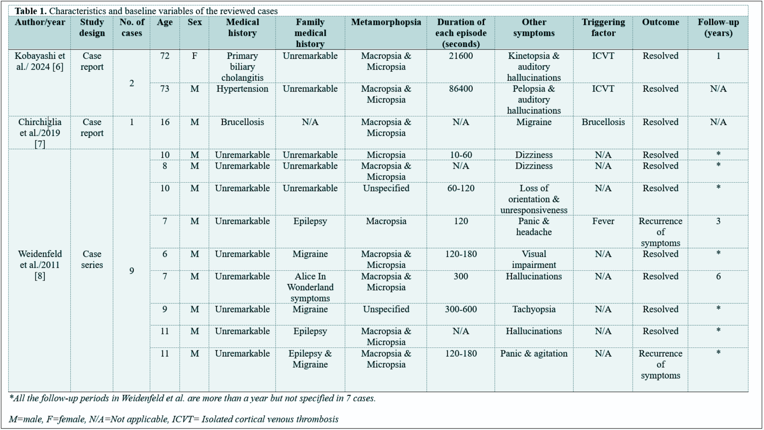
Unusual Presentation of Alice in Wonderland Syndrome: A Case Report and Literature Review
Yadgar N. Abbas, Meer M. Abdulkarim, Mohammed Q. Mustafa, Diyar A. Omar, Kayhan A. Najar, Karokh...
Abstract
Introduction
Alice in Wonderland Syndrome (AIWS) is an uncommon and frequently overlooked neuropsychiatric condition, marked by brief episodes of altered visual and somatosensory perception. This report presents a case of AIWS, highlighting the disorder's unusual nature.
Case presentation
A 21-year-old female sought evaluation due to episodic visual distortions and altered body perceptions lasting for six months, often accompanied by migraines. These episodes, including micropsia, macropsia, and derealization, typically occurred multiple times a week and lasted several minutes, with no clear triggers but worsening with stress or irregular sleep. She had a history of similar, less intense episodes in childhood. Neurological examination, MRI, EEG, and blood tests were all normal. She was diagnosed with AIWS related to her migraines and was prescribed propranolol, a stress management strategy, regular sleep, and cognitive behavioral therapy.
Literature review
Sixteen cases of AWIS were reviewed, of which only one had a family history of the condition. All of them experienced perceptual distortions, with macropsia and micropsia together appearing in eight cases, and hallucinations were present in four. Duration of symptoms ranged from one minute to one day. The triggering factors included Isolated cortical venous thrombosis and brucellosis. Treatments included lacosamide and paroxetine. Recurrence of symptoms was recorded in two patients.
Conclusion
Alice in Wonderland Syndrome is mostly a benign condition that can be resolved spontaneously or treated according to its associated cause; propranolol, improved sleep schedule, and cognitive behavioral therapy might yield good outcomes.
Introduction
Alice in Wonderland Syndrome (AIWS), first identified in 1955, is a rare perceptual disorder that causes significant distortions in how individuals perceive visual stimuli (metamorphopsias), their body image, and even the passage of time. Those affected may experience sensations such as seeing objects appear much larger or smaller than they are or feeling that their body is changing size [1]. The name of the syndrome is derived from Lewis Carroll's famous 19th-century novel Alice's Adventures in Wonderland, where the protagonist, Alice, experiences similar surreal phenomena, such as her body growing and shrinking, as well as other bizarre and disorienting occurrences. This condition is often described as a disruption in the brain's ability to process sensory input, leading to a distorted reality that can be confusing and disconcerting for the person experiencing it [2]. Although AIWS is rarely diagnosed, its actual prevalence may be underestimated, largely because clinicians often lack awareness of the condition, and patients, especially children, may struggle to describe their symptoms [3]. This condition is more commonly observed in younger individuals than in older adults [4]. After remaining relatively unnoticed for several decades, AIWS has recently started to gain scientific attention. This renewed focus is partly due to the advancement of functional imaging techniques, which now allow researchers to explore the brain's networks that mediate the disorder's symptoms [2,4]. Initially, AIWS was described in patients suffering from migraines and/or epilepsy and later found to be associated with temporoparietal lesions. It was also linked to psychiatric conditions such as depression and schizophrenia, as well as hypnagogic states and the use of various drugs, including hallucinogens and anticonvulsants [2]. Studies suggest that AIWS is usually a benign, self-limiting condition that resolves without the need for treatment [5].
This study aims to present a case of AIWS in a young adult with a history of migraines, emphasizing the outlandish and unusual nature of this disorder. The report was written following the CaReL guidelines, and only reliable, peer-reviewed sources were included while excluding any unreliable references or data [6,7].
Case presentation
Patient information
A 21-year-old female presented herself for evaluation at the suggestion of her family. She had experienced episodic visual distortions and disturbances in her perception of her body for six months. During these episodes, she said, her hands and feet appeared too large or too small, and the size and distance of everything around her seemed distorted. These episodes typically lasted several minutes and occurred multiple times a week, often coinciding with migraines. She was single and had sought help due to the frequency and disruption caused by these unusual perceptions.
The patient’s symptoms began approximately six months prior, without a clear trigger, involving visual distortions such as micropsia and macropsia, somesthetic distortions, and derealization. These episodes often coincided with migraines, characterized by unilateral headaches, photophobia, phonophobia, and nausea, resolving after several hours. The patient had no known triggers, though episodes were more frequent during stress or irregular sleep. Similar, less intense episodes occurred in childhood but were resolved by adolescence. The recent resurgence caused significant distress, prompting the patient to seek help.
The patient had no history of psychiatric illness, hospitalizations, or treatment, and had never used psychiatric medications. She was born at term with an unremarkable birth and past medical history, achieving all developmental milestones on time, with no early childhood illnesses or delays. She had a history of mild asthma treated with as-needed inhalers. There was no past head trauma, seizures, or infections, and the patient was healthy with no prior surgeries, and she also denied using alcohol, tobacco, or recreational drugs.
Mental state examination (MSE)
She was well-groomed and appropriately dressed. During the interview, she was cooperative and engaged, with no evidence of agitation or retardation. She was clear and coherent in her speech. With an anxious but congruent affect, she described feeling anxious due to the unpredictability of her symptoms. Her thought process was organized, goal-directed, and there was no evidence of thought disorder, delusions, or hallucinations. She described visual and somesthetic distortions consistent with Alice in Wonderland Syndrome, with no auditory hallucinations. Abounded with anxiety, but was eventually able to attend this socialization event. She exhibited good insight into her condition and preserved judgment.
Diagnostic investigation
The tests included MRI, EEG, and blood tests, all showing no abnormalities. Visual field testing was also normal, ruling out ocular causes for her symptoms. Given the normal test results, the diagnosis of AIWS was made based on the patient's reported symptoms and history of migraines.
Management
She was placed on prophylactic treatment with propranolol, a beta-blocker, to control her migraines. She was also advised to manage stress and maintain a consistent sleep schedule to reduce the likelihood of migraines. In addition, cognitive behavioral therapy was implemented to help combat the distress associated with her episodes.
Follow-up
After several months, both the frequency and intensity of her migraines diminished, and the severity of her AIWS episodes also lessened. Nonetheless, she continued to experience occasional perceptual distortions.
Discussion
In the existing literature, AIWS is typically described as a consequence of conditions like migraines, without leading to irreversible brain damage. However, infections have become the most commonly reported cause linked to AIWS cases, especially in children, surpassing migraines and epilepsy. Because of this, the exact nature of the lesions and the underlying pathology of AIWS remain difficult to identify and poorly understood [2,8].
AIWS symptoms have expanded to include 42 visual symptoms and 16 somesthetic and other nonvisual symptoms [1]. Individuals with AIWS experience changes in visual perception, such as micropsia (where objects appear smaller), macropsia (where objects appear larger), teleopsia (where objects seem farther away), and pelopsia (where objects seem closer). Similar to the reported case, all reviewed patients experienced perceptual distortions. Macropsia and micropsia occurred concurrently in nine cases, while hallucinations were reported in five. Incoherent speech, anxiety, and derealization were also noted as accompanying features of the visual distortions (Table 1) [3,4, 8-12].
Author/year |
No. of cases |
Age/Sex |
Medical history |
Family medical history |
Metamorphopsia |
Duration of each episode (minutes) |
Other symptoms |
Triggering factor |
Treatment |
Outcome |
Follow-up (years) |
Alexis Demas/2025 [12] |
1 |
68/M |
Hypertension |
Unremarkable |
Micropsia |
N/A |
Incoherent speech, akinetic-rigid Parkinsonian syndrome, apathy, anxiety, and delusional thoughts |
N/A |
Levetiracetam |
Death |
0.16 |
Ansari et al./2025 [3] |
1 |
10/F |
Unremarkable |
Unremarkable |
Macrosomatognosia & Micropsia |
10-20 |
Time distortion & derealization |
N/A |
Supportive care & reassurance |
Resolved |
0.5 |
Ahmed et al./2025 [4] |
1 |
94/F |
Type 2 diabetes, hypertension, hypothyroidism, GERD & anxiety |
Unremarkable |
Macropsia |
N/A |
Visual & auditory hallucinations |
Ischemic stroke |
Risperidone, olanzapine & Quetiapine |
Improvement in symptoms |
N/A |
Mbizvo et al./2025 [11] |
1 |
69/M |
Ischemic stroke, caecal cancer, hypertension & type 2 diabetes |
Unremarkable |
Macropsia & Micropsia |
N/A |
Difficulties with color perception |
Ischemic stroke |
phenytoin |
Death |
N/A |
Kobayashi et al./ 2024 [8] |
2 |
72/F |
Primary biliary cholangitis |
Unremarkable |
Macropsia & Micropsia |
360 |
Kinetopsia & auditory hallucinations |
ICVT |
Lacosamide |
Resolved |
1 |
73/M |
Hypertension |
Unremarkable |
Macropsia & Micropsia |
1440 |
Pelopsia & auditory hallucinations |
ICVT |
N/A |
Resolved |
N/A |
||
Chirchiglia et al./2019 [9] |
1 |
16/M |
Brucellosis |
N/A |
Macropsia & Micropsia |
N/A |
Migraine |
Brucellosis |
Oral paroxetine |
Resolved |
N/A |
Weidenfeld et al./2011 [10] |
9 |
10/M |
Unremarkable |
Unremarkable |
Micropsia |
0.16-1 |
Dizziness |
N/A |
Unspecified |
Resolved |
* |
8/M |
Unremarkable |
Unremarkable |
Macropsia & Micropsia |
N/A |
Dizziness |
N/A |
Unspecified |
Resolved |
* |
||
10/M |
Unremarkable |
Unremarkable |
Unspecified |
1-2 |
Loss of orientation & unresponsiveness |
N/A |
Unspecified |
Resolved |
* |
||
7/M |
Unremarkable |
Epilepsy |
Macropsia |
2 |
Panic & headache |
Fever |
Unspecified |
Recurrence of symptoms |
3 |
||
6/M |
Unremarkable |
Migraine |
Macropsia & Micropsia |
2-3 |
Visual impairment |
N/A |
Unspecified |
Resolved |
* |
||
7/M |
Unremarkable |
Alice In Wonderland symptoms |
Macropsia & Micropsia |
5 |
Hallucinations |
N/A |
Unspecified |
Resolved |
6 |
||
9/M |
Unremarkable |
Migraine |
Unspecified |
5-10 |
Tachyopsia
|
N/A |
Unspecified |
Resolved |
* |
||
11/M |
Unremarkable |
Epilepsy |
Macropsia & Micropsia |
N/A |
Hallucinations |
N/A |
Unspecified |
Resolved |
* |
||
11/M |
Unremarkable
|
Epilepsy & Migraine |
Macropsia & Micropsia |
2-3 |
Panic & agitation |
N/A |
Unspecified |
Recurrence of symptoms |
* |
||
*All the follow-up periods in Weidenfeld et al. are more than a year, but not specified in 7 cases. M: male, F: female, N/A: Not applicable, ICVT: Isolated cortical venous thrombosis, GERD: Gastroesophageal reflux disease. |
Additionally, some patients with AIWS report abnormal changes in their perception of time, experiencing it as either speeding up or slowing down [2]. Distortions in the perception of time were present in the form of tachyopsia (environment moving or changing at an abnormally fast rate) in one of the reviewed cases [10]. Notably, a patient with AIWS is always aware that these distorted perceptions are not real, which distinguishes AIWS from conditions like psychosis, where the patient may perceive such distortions as part of their reality [2]. In AIWS, symptom duration is usually quite short, with each episode varying from less than 5 min to at most 30 min; however, 2 of the reviewed cases had episodes lasting up to 6 and 24 hours, adding another angle of uncertainty to this condition [8].
Kobayashi et al. reported 2 cases of AWIS, with lesions located in the right occipital lobe. To date, there have been about 10 cases of AWIS with bilateral or unilateral occipital lobe lesions [8]. While this finding suggests that occipital lesions may play a role in the development of AIWS, the majority of the cases do not involve brain damage. Similar to the reported case, 12 of the reviewed cases had no brain damage [3, 9-12]. Although several brain networks related to visual perception have been proposed as contributors to AIWS symptoms, imaging studies have not determined a clear cause, and some reports present conflicting results. However, the high number of reports linking migraine and epilepsy with AIWS suggests that an incomplete or widespread brain dysfunction may play a role in the development of the disorder [6].
Recent reports suggest that infectious diseases, particularly among children, are a leading cause of this Syndrome. These include Epstein-Barr virus, cytomegalovirus, Coxsackie virus B1, and varicella-zoster virus. The syndrome has also been linked to bacterial infections like Mycoplasma pneumoniae, Borrelia burgdorferi, and Streptococcus pyogenes, as well as protozoan and prion infections [2]. A case of AWIS associated with brucellosis was among the reviewed cases, adding to the increased infection-related cases [9].
Since 1955, fewer than 200 cases of AIWS have been recorded. However, the literature indicates that this may only represent a small fraction, as up to 30% of adolescents in the general population experience some of the individual symptoms of AIWS, albeit occasionally and briefly. [1]. Since it is not included in major classifications such as the ICD-10 or DSM-5, diagnosing AIWS depends on careful history-taking, a comprehensive physical examination (including neurologic and often otologic or ophthalmic assessments), and a good understanding of its diverse symptoms and possible causes. If a central origin is suspected, further tests like blood tests, EEG, and brain MRI are recommended, though the likelihood of identifying any observable lesions is generally considered low, as seen in the present case [1].
No specific treatment exists for AIWS; management generally focuses on addressing the underlying cause. Among the reviewed cases, interventions included beta-blockers, antipsychotics, antiepileptics, SSRIs, and supportive care, with symptoms resolving in 12 patients and recurrence noted in two [3,4,8-12]. Research indicates that antipsychotics are infrequently used in AIWS, and their effectiveness is generally considered limited. Moreover, when perceptual distortions occur alongside psychotic symptoms, it is important to note that antipsychotics, such as risperidone, may occasionally precipitate or exacerbate these distortions by lowering the threshold for epileptic activity [4].
Conclusion
AIWS is mostly a benign condition that can be resolved spontaneously or treated according to its associated cause; propranolol, improved sleep schedule, and cognitive behavioral therapy might yield good outcomes.
Declarations
Conflicts of interest: The authors have no conflicts of interest to disclose.
Ethical approval: Not applicable.
Patient consent (participation and publication): Written informed consent was obtained from the patient for publication.
Funding: The present study received no financial support.
Acknowledgements: None to be declared.
Authors' contributions: YNA was a significant contributor to the conception of the study and the literature search for related studies. MMA was involved in the literature review, study design, and manuscript writing. MQM, DAO, KAN, KKM, and SSA were involved in the literature review, the study's design, and the critical revision of the manuscript, and they participated in data collection. YNA and MMA confirm the authenticity of all the raw data. All authors approved the final version of the manuscript.
Use of AI: ChatGPT-3.5 was used to assist in language editing and improving the clarity of the manuscript. All content was reviewed and verified by the authors. Authors are fully responsible for the entire content of their manuscript.
Data availability statement: Not applicable.

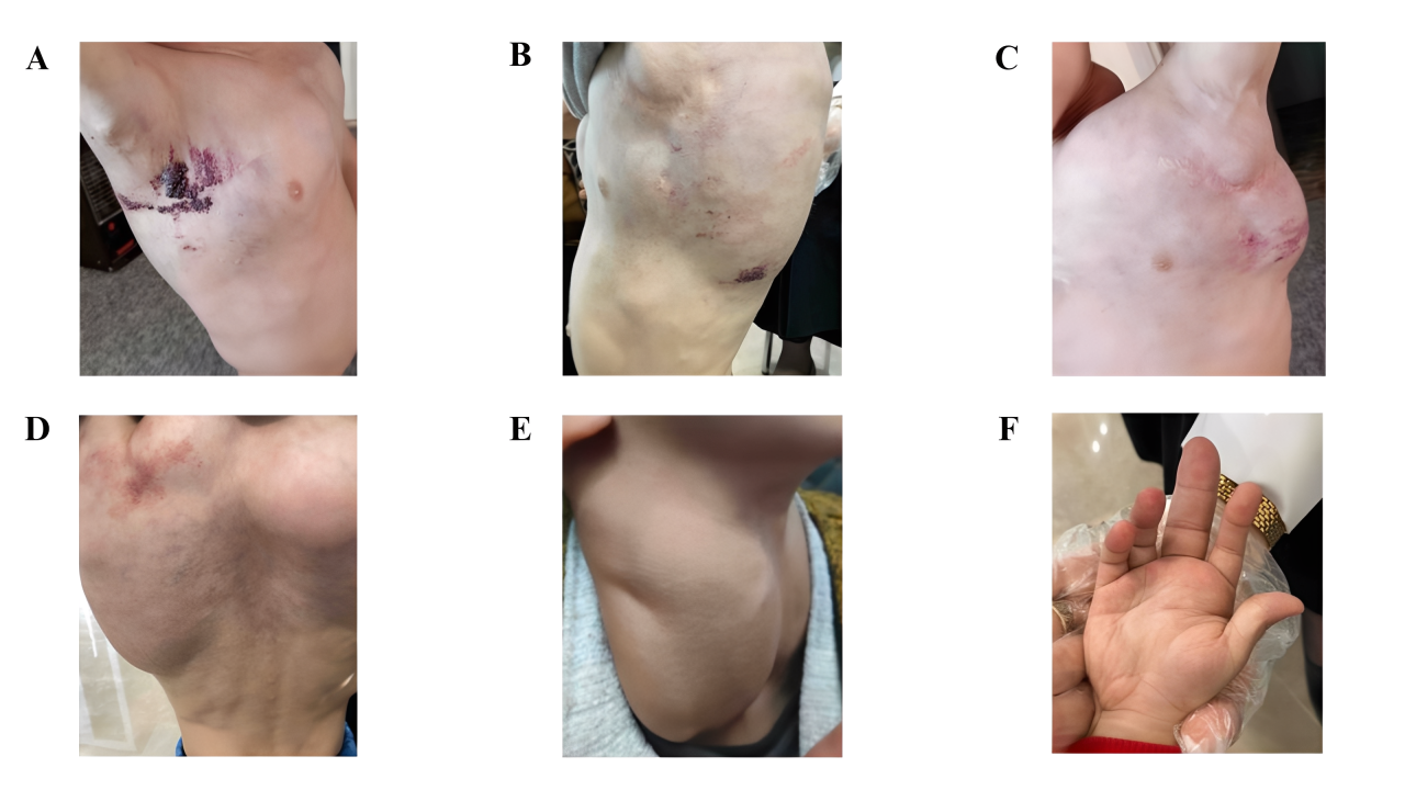
Unusual Presentation of Mixed Lymphatic Malformation: A Case Report with Literature Review
Ronak S. Ahmed, Shvan O. Siddiq, Lawen J. Mustafa, Omed M. Hussein, Sakar O. Arif, Dana HB....
Abstract
Introduction
There is a scarcity of reports on mixed lymphatic malformation. This case highlights a child with an extensive mixed lymphatic malformation, disfiguring multiple parts of the body.
Case presentation
A 3.5-year-old boy presented with multiple vesicular “frog-spawn” lesions that affected the right mid-axillary region, left axilla, and left upper back. The patient also had marked bilateral cervical swelling, extending laterally on both sides, with prominent bulges measuring approximately up to 10 cm that extended to involve the entire right upper limb dawn to the hand. Bilateral axillary swelling was observed. Additionally, swelling was evident in the entire middle and upper back, with a pronounced rounded bulge in the right upper back. Surgical intervention of the left axillary mass was performed, which was consistent with macrocystis lymphatic malformation on histopathology, but the condition recurred during follow-up.
Literature review
Few cases of microcystic lymphatic malformation with extensive deep lymphatic involvement have been reported in the literature. There was a female predilection with reported involvement of the forearm, thigh, vulva, axilla, jaw, and even the tongue. The patients' ages ranged from infancy to adulthood, with some patients complaining of skin lesions and recurrent cellulitis for years, sometimes leading to sepsis and death. There is variable outcome for surgical intervention in such cases.
Conclusion
Mixed lymphatic malformation can present extensively affecting several parts of the body causing significant disfigurement with possible recurrence after surgical intervention.
Introduction
The lymphatic system is essential for regulating plasma volume and preventing tissue pressure buildup, as it reabsorbs a significant portion of the fluid and proteins that leak out the blood vasculature. Additionally, it supports cell nutrition and aids the immune response by directing antigen-presenting cells to lymph nodes. In the skin, lymphatic vessels serve as a critical exit route for Langerhans cells [1]. Anomalies in the development of lymphatic channels can lead to various diseases, including lymphatic malformation which can be either localized or diffuse and may be either congenital or acquired [2]. Notably, lymphatic malformation shows no variation across racial groups, with up to 90% of cases diagnosed before the age of two [3].
The International Society for the Study of Vascular Anomalies (ISSVA) established a comprehensive classification system that categorizes vascular anomalies based on their clinical characteristics and whether they are accompanied by other symptoms, which includes cystic lymphatic malformation, Kaposiform lymphangiomatosis, Gorham–Stout disease, and Channel-type lymphangioma. The cystic type is further categorized into macrocystic lymphatic malformation (diameter greater than 1cm), microcystic lymphatic malformation (diameter less than 1cm), formerly called lymphangioma circumscriptum, and mixed cystic lymphatic malformation. Histologically, the lymphatic cysts may either appear empty or contain a protein-rich fluid abundant in lymphocytes and macrophages [3].
Cutaneous lymphatic malformation represents only 4% of all vascular tumors. Microcystic lymphatic malformation is an uncommon superficial variant that primarily affects the axilla, groin, thighs, and buttocks. The “frog-spawn” appearance is due to the distinguished formation of dispersed clusters of small, thin-walled, translucent vesicles on the skin. Additionally, microcystic lymphatic malformation may contain a prominent vascular or hemorrhagic component, causing the lesion's color to vary from pink to red to black, depending on the proportion of blood to lymph within the vesicles. The present report aims to describe a case of mixed lymphatic malformation with extensive involvement, disfiguring various parts of the body [4].
The current study was written in line with CaReL guidelines and the credibility of references has been verified using Kscien’s list [5,6].
Case Presentation
Patient information
A 3.5-year-old male presented with complaints of intermittent bleeding from skin lesions in the right axillary region. Additionally, the patient complained of extensive swelling in the neck, axilla, and upper back, and swelling of the right middle finger. There was no family history of similar skin lesions or swelling. The patient's past surgical history was notable for herniorrhaphy and axillary mass resection.
Physical examination
On physical examination, there were multiple skin-colored compressible masses on the neck, axillae, trunk, and upper limbs. On the axillae and upper back, there were multiple small groups of papulovesicular lesions with characteristic “frog-spawn” appearance with many bleeding points. The patient had marked bilateral cervical swelling, extending laterally on both sides of the neck, with prominent bulges measuring approximately up to 10 cm on each side, that extended to involve the entire right upper limb down to the hand. Bilateral axillary swelling was observed, accompanied by a scar in the left axilla, indicative of previous surgical intervention. Additionally, swelling was evident in the entire middle and upper back, with a pronounced rounded bulge in the right upper back. The swelling extended across the back, with slight marbling noted on the overlying skin. There was also swelling of the chest, most prominently around the sternum. In addition, there was swelling in the right middle finger (Figure 1).
Diagnostic approach
The skin lesions were diagnosed as microcystic lymphatic malformation (lymphangioma circumscriptum) based on their location, the characteristic appearance of the vesicular lesions, and the history of oozing blood. The involvement of deeper lymphatic system swelling further supported the diagnosis.
The laboratory blood investigations including complete blood count, thyroid stimulating hormone, C-reactive protein, and D-dimer were all within the reference range. Ultrasonography of the axilla and inguinal regions showed fairly symmetric, bilateral axillary soft tissue lesions with ill-defined, heterogeneous echogenicity, measuring approximately 8×4 cm. Small cystic areas were present within the lesions, with minimal internal vascularity but no evidence of phleboliths or prominent vessels. The sonographic features were non-specific, and potential differentials included hibernoma, lipoblastoma, and atypical hemangioma. A left axillary lymph node measuring 13×8 mm was noted; however, the bulk of the swelling was attributed to the soft tissue mass. No enlarged inguinal lymph nodes were observed, but a bilateral large inguinal hydrocele was present, along with bilateral undescended testes located in the inguinal regions. The other abdominal organs showed no abnormalities.
Contrast-enhanced computed tomography scan of the neck and upper chest showed normal lung parenchyma and a non-enhancing hypodense mass in the left axilla extending to the left supraclavicular region and a hypodense mass in the right side of the neck (Figure 2). The axillary magnetic resonance imaging (MRI) revealed bilateral symmetrical axillary masses, measuring 6×5×5 cm on the right and 6.5×5×4.5 cm on the left. These masses exhibited homogeneous high signal intensity on both T1 and T2-weighted images and were suppressed on fat-saturated sequences, with no enhancement post-contrast. The masses were associated with multiple enlarged axillary lymph nodes, the largest measuring 13×10 mm on the right and 13×9 mm on the left. Lipoblastoma was suggested as a differential, and biopsy was advised.
Biopsy of the axillary masses showed multiple fatty gray and tan pieces of tissue, the largest one measuring 85x80x40 mm. The microscopic sections showed multiple dilated cavernous and capillary size vascular spaces lined a by single layer of bland endothelium with flattened nuclei, surrounded by nodular aggregates of small mature lymphocytes and embedded in large lobules of mature adipose tissue, with areas that contain fetal-type skeletal muscle in process of maturation. There was no evidence of granulomata or invasive malignancy in the examined sections. Histopathology results were most consistent with macrocystic lymphatic malformation. A diagnosis of mixed lymphatic malformation was made.
Therapeutic intervention and follow-up
The patient previously underwent surgery to address the left axillary swelling; however, the lymphangioma recurred without significant improvement. Once-daily administration of sildenafil 10 mg and propranolol 10 mg yielded a good clinical response within the first three months, however on follow up there was an increase in the size of the mass. The frequency sildenafil dose was increased to 10 mg twice daily, and Pulsed Dye Laser (PDL) was applied for the microcystic lymphatic lesions for three sessions with no satisfactory response. At present, the patient is under observation with regular follow-up to monitor any potential complications or changes in the swellings that may require further intervention.
Discussion
The human skin comprises two distinct lymphatic plexuses. The superficial plexus, located near the subpapillary arterial network, consists of thin, non-valvular vessels extending into the dermal papillae. Lymph from this network drains into larger vessels situated in the lower dermis and the superficial subcutaneous tissue. The deep lymphatic plexus, positioned beneath the second arterial network, contains numerous valves and closely parallels the venous collecting system of the lower dermis [1].
The underlying pathophysiology of lymphatic malformation is characterized by fluid-filled muscular cisterns situated within the subcutaneous tissue, which establish a connection with the surface through dilated dermal lymphatic channels. Cutaneous involvement arises as a secondary consequence of the pressure exerted within the cisterns, driven by the pulsatile activity of their muscular walls. These cisterns do not communicate with the normal lymphatic system and lack any functional physiological purpose. The full extent of deep lymphangiomatosis cannot be accurately evaluated through physical examination alone, as the cisterns may extend significantly beyond the region occupied by the superficial vesicles [7]. For instance, a 13-year-old female reported by Palmer et al. complained of superficial lesions affecting the right inner thigh diagnosed as microcystic lymphangioma, but the right thigh had a 50cm thickness compared to the left side of 47.5cm. Further imaging revealed atypical expansion of the lymphatic channel stretching from the femoral region to the right para-aortic area [8]. Furthermore, a 15-year-old had more than 5 years of oozing from the microcystic lymphatic malformation affecting the left anteromedial inner thigh with deeper lymphatic involvement reaching the labia majora causing significant swelling and difficulty ambulating and disfigurement, during the literature review more cases have been identified with various parts of the body being affected (Table 1) [ 7-11].
Author/ year of publication |
Age (years) |
Sex |
Duration of complaint |
Microcystic lymphangioma location |
Deeper lymphatic involvement |
Diagnostic imaging |
Histopathology |
Treatment |
Follow-up |
Outcome |
Palmer et al./ 1978 [8] |
13 |
F |
Several years |
Right inner thigh |
Right thigh enlargement |
A lymphangiogram demonstrated abnormal dilation of the lymphatic channel extending from the femoral region up to the right para-aortic region |
Dilated endothelial lined lymph channels |
N/A |
N/A |
N/A |
Anees et al./ 2006 [9] |
15 |
F |
Six years |
Left inner thigh |
From the left groin up to the proximal half of left thigh, localized on the medial aspect, and involving the left labia majus |
N/A |
Dilated, large “cistern” of lymph vessels lined by a single layer of endothelium and extending to the deeper zone of the dermis. |
Surgical intervention |
1.5 years |
No recurrence |
Mordehai et al./ 1998 [10] |
1 month |
M |
Since birth |
Right forearm |
Right axilla |
Chest radiograph showing dilatation of upper mediastinum, right side
CT scan of chest showing cystic mass 3 cm in diameter in anterior mediastinum adherent to superior vena cava on right side of trachea |
Confirmatory of lymphangioma circumscriptum |
Wide excision of the axillary lymphangioma, right thoracotomy. |
9 years |
recurrent episodes of cellulitis of the right forearm and axilla with local recurrence of the lymphangioma in the form of small vesicles 1±2 mm in diameter along the scar tissue of previous suture lines and at the border of the skin grafts and lymphorrhea |
Rao et al./ 1998 [11] |
25 |
F |
Since birth |
Right temporal area, raised lesions over the right half of the tongue |
Right jaw |
N/A |
multiple dilated, cystic vessels in the papillary and sub-papillary dermis, containing lymph and occasional erythrocytes and were lined by a single layer of endothelium |
N/A |
N/A |
N/A |
Beard et al./ 1995 [7] |
9 |
F |
Since birth |
Buttocks and the stump of her right leg in the area of previous amputation |
Entire right lower extremity, extensive abdominopelvic lymphangiomatosis |
Radiographs revealed a soft tissue swelling without evidence of bony enlargement. lymphangiogram were most consistent with a lymphangiohemangioma |
Biopsy was consistent with lymphangioma circumscriptum |
amputation above the right knee at age four. Radiotherapy. frequent scissor excisions and cryosurgery. carbon dioxide laser ablation of the coccygeal, intergluteal, buttock, perineal, and perianal lymphangiomatosis |
Until the age of 15 |
Recurrent cellulitis, temporary improvement after interventions. The patient ultimately died at 15 after cellulitis, UTI, and resultant sepsis. |
The skin features of microcystic lymphangioma make its diagnosis straightforward but it can be misdiagnosed as other conditions, with a broad list of differential diagnoses including dermatitis herpetiformis, angiokeratoma circumscriptum, herpes simplex, herpes zoster, and hemangioma. In an interesting case by Juca et al, due to the verrucous appearance of the microcytic lymphangioma affecting a toe, leishmaniasis, skin tuberculosis, and warts were suspected first. However, histopathology was suggestive of microcystic lymphatic malformation showing convoluted ectatic lymph vessels in the papillary dermis [12]. Histopathology of the current patient revealed dilated vascular spaces lined by flat endothelium, surrounded by lymphoid aggregates, mature fat, and maturing fetal-type skeletal muscle.
Although lymphatic malformations are benign and typically a cosmetic concern, they can cause symptoms like burning, itching, and pain especially when complicated by infection, hemorrhage, or mechanical irritation. Leakage of blood-tinged, sometimes foul-smelling lymphatic fluid, either spontaneously or after minor trauma, can lead to discomfort and embarrassment. Open vesicles may also increase the risk of infection, leading to recurrent cellulitis and sometimes leading to sepsis and death. Due to cosmetic concerns, emotional distress, or frequent infections, patients often seek intervention [4,7]. Previously lymphangiogram was used to assess the extent of deeper lymphatic involvement as was the case by Palmer et al and Beard et al. Currently, computed tomography and magnetic resonance imaging are utilized more often [7-9]. For example, in the case report by Mordehai et al. computed tomography of a 1-month-old was shown to have a 3 cm cystic mass located in the anterior mediastinum, adjacent to the right side of the trachea and adherent to the superior vena cava. While on physical examination there was a group of vesicles on the right forearm with deeper cystic hygroma causing significant swelling of the right axilla. Interestingly, when wide excision of the axillary lymphangioma was carried out, the microcystic lymphatic malformation was then present at the site of the prior surgical site and suture lines with repeated episodes of cellulitis and lymphorrhea during the nine-year follow-up period [10]. Similarly, the current case had excision of the lymphangioma in the left axilla however, there was no recurrent cellulitis or vesicle formation at the scar site. The surgery was deemed ineffective as the deeper lymphatic involvement of the axilla recurred. Nonetheless, Surgical excision remains the primary treatment modality for the extensive/classical subtype, despite the potential for recurrence, as the surgical approach is predicated on the complete excision of all sequestrated lymphatic cisterns within the subcutaneous plane, as they are the principal etiological factor. The patient reported by Anees et al. resulted in good outcomes with no sign of recurrence 1.5 years after the operation of the medial left thigh. Other treatment modalities include intra-lesional sclerotherapy with doxycycline or Picibanil, as well as vaporization using carbon dioxide laser [9]. In a systematic review including 28 individuals with carbon dioxide laser ablation of microcystic lymphangioma, eight of them were disease-free and ten of them showed partial recurrence up to a three-year follow-up period, serving as an effective therapeutic option with a tolerable side-effect profile. Notably, it may also outperform surgical intervention for lesions with extensive surface area and deep involvement, though additional research is needed to confirm this advantage [4].
One limitation of our study is the absence of histopathological images, as only the histopathology reports were available. In addition, the relatively short follow-up period does not showcase the long-term physical and psychological outcomes of extensive mixed lymphatic malformation affecting several parts of the body.
Conclusion
Mixed lymphatic malformation can present extensively affecting several parts of the body causing significant disfigurement with possible recurrence after surgical intervention.
Declarations
Conflicts of interest: The authors have no conflicts of interest to disclose.
Ethical approval: Not applicable.
Patient consent (participation and publication): Written informed consent was obtained from the patient for publication.
Funding: The present study received no financial support.
Acknowledgements: None to be declared.
Authors' contributions: RSA and SOS were significant contributors to the conception of the study and the literature search for related studies. DH and JIH were involved in the literature review, study design, and manuscript writing. LJM, OMH, SOA, DHBMS and FJS were involved in the literature review, the study's design, and the critical revision of the manuscript, and they participated in data collection. RSA and DH confirm the authenticity of all the raw data. All authors approved the final version of the manuscript.
Use of AI: ChatGPT-3.5 was used to assist in language editing and improving the clarity of the manuscript. All content was reviewed and verified by the authors. Authors are fully responsible for the entire content of their manuscript.
Data availability statement: Not applicable.

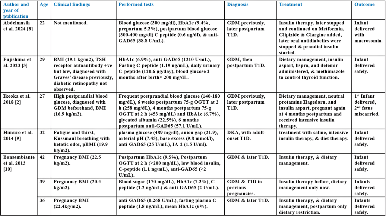
Pregnancy and Challenging Transient Anti-GAD65 Positivity: A Case Report with Literature Review
Shaho F. Ahmed, Sharaza Q. Omer, Rawezh Q. Salih, Huda M. Muhammad, Nahida H.A Ahmed, Jamal M....
Abstract
Introduction
During pregnancy, women may develop blood glucose abnormalities like gestational diabetes mellitus (GDM) or, rarely, type 1 diabetes (T1D), which can lead to complications. Anti-GAD65 is a key antibody used in diagnosing T1D. This study presents a rare case of T1D developing a week before birth, with transient anti-GAD65 positivity.
Case presentation
A 38-year-old patient who delivered a baby a week earlier and had high blood glucose was admitted to the hospital with shortness of breath, chest tightness, abdominal pain, generalized weakness, nausea, and repeated vomiting. She had elevated anti-GAD65 and was diagnosed with T1D and diabetic ketoacidosis. Insulin injection was prescribed, with lifestyle modifications, later oral hypoglycemic medications added. After a few months, both anti-IA2 and anti-GAD65 antibodies were negative.
Literature review
Seven cases of T1D during or after pregnancy were reviewed. Six reported BMI. The mean HbA1c was >7.63%. Mean anti-GAD65 was 190.74 U/mL, two were borderline and one negative. Six had previously diagnosed GDM. Treatments varied, including insulin and dietary management. All infants were safely delivered, one miscarried in a subsequent pregnancy. Insulin resistance increases during pregnancy due to hormonal changes, raising the risk of GDM, T1D, and type 2 diabetes. Emerging postpartum, often indicated by anti-GAD65 antibodies, though levels can fluctuate. Cases show complications like preeclampsia, DKA, and miscarriage. Early detection, strict glucose control, and monitoring antibody patterns are critical for managing risks and improving maternal and fetal outcomes.
Conclusion
Blood glucose should be monitored during pregnancy, and anti-GAD65 may signal T1D, requiring appropriate management.
Introduction
Type 1 diabetes (T1D) is a condition characterized by a lack of the hormone insulin due to an autoimmune response that destroys the pancreas's insulin-producing β cells [1]. During pregnancy, women may develop an abnormal tolerance to glucose, which is not severe enough to be classified as overt diabetes mellitus, known as gestational diabetes mellitus (GDM). This condition increases the possibility of type 2 diabetes (T2D) after birth, and the risk of T1D, especially if they have autoantibodies targeting the beta cells of the pancreas [2]. Around 2-17% of expectant mothers empierce GDM. While glucose metabolism typically returns to normal after childbirth, there is a high chance, between 34% up to half of the cases, that it will recur in future pregnancies, with around 6% developing into T1D [3]. Throughout an asymptomatic period of variable length, different autoantibodies targeting islet cell autoantigens can be identified, and these play a key role in recognizing individuals at an increased risk of developing clinical disease. Glutamic acid decarboxylase (GAD) is a key enzyme, antibodies against said enzyme or anti-GAD65 are one of the primary antibodies associated with T1D, with two isoforms having molecular weights of 65 kDa and 67 kDa. This antibody is found in most individuals with preclinical or recently diagnosed T1D. [4]. It is well established that pregnancies that involve T1D face a greater risk of complications affecting the mother, fetus, and newborn compared to those with uncomplicated pregnancies [5]. Poorly managed high blood glucose during pregnancy can result in serious complications, including stillbirth, miscarriage, congenital disabilities, and pregnancy-related conditions like hypertension or preeclampsia [1]. The aim of this study is to show a rare case of T1D developing a week before giving birth, with a transient anti-GAD65 positive test result, and a literature review. The references used in this case report have been checked for reliability and compiled with CaReL guidelines [6,7].
Case Presentation
Patient information
A 38-year-old female was admitted to the hospital with shortness of breath, chest tightness, abdominal pain, generalized weakness, nausea, and repeated vomiting that had persisted for two days before admission. She had been pregnant and had delivered a stillborn baby one week earlier at 39 weeks of gestation.
Clinical findings
On clinical examination, the patient appeared distressed and exhibited tachypnoea. Her heart rate was 110 beats per minute, respiratory rate was 32 cycles per minute, blood pressure was 110/70 mmHg, and her temperature was 38°C. Her BMI was 34 kg/m². She had no history of chronic disease.
Diagnostic approach
The patient was admitted to the hospital, resuscitated, and diagnosed with diabetic ketoacidosis (DKA), based on the presence of ketone bodies in the urine (3+) and acidosis confirmed by arterial blood gas analysis (pH 7.16, bicarbonate 12 mmol/L), her blood glucose was over 300 mg/dL. Anti-GAD test came back elevated at (65.1 U/mL), leading to a diagnosis of T1D.
Therapeutic intervention
Several days before her scheduled Cesarean section, she was admitted to the hospital due to elevated blood pressure and blood glucose levels. She was treated with insulin therapy to manage her condition. Following the surgery, the patient was evaluated by an endocrinologist for her hyperglycemic condition. Based on her medical history, the decision was made to discontinue insulin therapy and initiate lifestyle modifications along with oral hypoglycemic medications. However, two days after this change in treatment, she developed DKA. To treat her DKA, she received intravenous insulin infusion and was rehydrated with normal saline and received potassium.
Follow-up
A few months after starting insulin therapy, she underwent testing for T1D-related autoantibodies, which came back within normal ranges. The results were negative for both anti-IA2 and anti-GAD65 antibodies.
Discussion
The sensitivity to insulin declines during pregnancy, primarily due to the influence of placental hormones. Human growth hormone, along with human placental lactogen, plays a key role in regulating maternal metabolism and supporting the growth of the fetus, with levels gradually increasing throughout the first and second trimesters. Pregnancy is also associated with a reduced secretion of human growth hormone in response to low blood glucose, which further contributes to the resistance toward insulin. These effects are compounded by hormonal changes involving progesterone, prolactin, cortisol, adiponectin, and leptin [1]. Seven cases of T1D arising during or after pregnancies have been reviewed (Table 1) [2, 3, 8-10]. Six cases mentioned the patient’s body mass index, one of them mentioning the pre-pregnancy body mass index. The mean HbA1c was over 7.63, only one case didn’t mention performing a HbA1c test. The mean anti-GAD65 was 190.74 U/mL, with two cases being borderline at around 2 U/mL and one negative at 0.268 U/mL. Six cases were associated with GDM before T1D. Different treatments and therapeutics were used, including insulin therapy and dietary managements. All the infant were delivered safely, but one case had another pregnancy a few months later and the fetus miscarried.
Author and year of publication |
Age |
Clinical findings |
Performed tests |
Diagnosis |
Treatment |
Outcome |
Abdelmasih et al. 2024 [8] |
22 |
Not mentioned. |
Blood glucose (300 mg/dl), HbA1c (9.4%, prepartum 5.3%), postpartum blood glucose (300-400 mg/dl) C peptide (0.6 ng/dl), & anti-GAD65 (38.8 U/mL). |
GDM previously, later postpartum T1D. |
Insulin therapy, later stopped and continued on Metformin, Glipizide & Glargine added, later oral antidiabetics were stopped & prandial insulin started. |
Infant delivered with macrosomia. |
Fujishima et al. 2023 [3] |
29 |
BMI (19.1 kg/m2), TSH receptor autoantibody +ve but low, diagnosed with Graves’ disease previously, diabetic retinopathy not observed. |
HbA1c (6.9%), anti-GAD65 (1210 U/mL), Fasting C-peptide (1.19 ng/mL), daily urinary C-peptide (120.6 μg/day), blood glucose 2 months after birth≥ 200 mg/dL. |
GDM, then postpartum T1D. |
Dietary management, insulin aspart, lispro, and detemir administered, & methimazole to control thyroid function. |
Infant delivered safely. |
Ikeoka et al. 2018 [2] |
27 |
High postprandial blood glucose, diagnosed with GDM beforehand, BMI (16.9 kg/m2). |
Frequent postprandial blood glucose (140-180 mg/dL), 6 weeks postpartum 75-g OGTT at 2 h (258 mg/dL), 4 months postpartum 75-g OGTT at 2 h (453 mg/dL) and HbA1c (6.7%), glycated albumin (22.5%), 6 months postpartum anti-GAD65 (57.1 U/mL). |
GDM previously, later postpartum T1D. |
Dietary management, neutral protamine Hagedorn, and insulin aspart, pregnant again at 4 months postpartum and received intensive insulin therapy. |
1st Infant delivered, 2nd fetus miscarried. |
Himuro et al. 2014 [9] |
32 |
Fatigue and thirst, Kussmaul breathing with ketotic odor, pBMI (19.9 kg/m2). |
plasma glucose (489 mg/dl), anion gap (21.9), arterial pH (7.45), base excess (9.8 mmol/l), anti-GAD65 (25 U/mL), IA-2 (1.5 U/ml). |
DKA, with adult-onset T1D. |
treatment with saline, intensive insulin therapy, & diet therapy. |
Infant delivered safely. |
Bonsembiante et al. 2013 [10] |
42 |
Pregnancy BMI (22.5 kg/m2). |
Postpartum HbA1c (9.5%), Postpartum OGTT at 2 h (>200 mg/dL), low blood insulin, C-peptide (1.1 ng/mL), anti-GAD65 (>2 U/mL). |
GDM & later T1D. |
Insulin therapy, & dietary management. |
Infant delivered safely. |
39 |
Pregnancy BMI (20.4 kg/m2). |
Blood sugar (170 mg/dL), HbA1c (7.3%), C-peptide (1.2 ng/mL) & anti-GAD65 (2 U/mL). |
GDM & T1D in previous pregnancies. |
Insulin therapy before, dietary management only now. |
Infants delivered safely. |
|
36 |
Pregnancy BMI (22.4kg/m2). |
anti-GAD65 (0.268 U/mL), fasting plasma C-peptide (1.8 ng/mL), mean HbA1c (6%). |
GDM & later T1D. |
Insulin therapy, & dietary management, postpartum only dietary restriction. |
Infants delivered safely. |
|
T1D = Type 1 diabetes, GDM = Gestational diabetes mellitus, +ve = Positive, pBMI = Pre-pregnancy body mass index, BMI = Body mass index, TSH = Thyroid-stimulating hormone, OGTT = Oral glucose tolerance test, HbA1c = Hemoglobin A1c, DKA = Diabetic ketoacidosis, IA-2 = Islet antigen-2, h = Hours. |
Pregnancies with T1D who have poor blood glucose control face a much higher risk of developing preeclampsia compared to those who do not. Even if T1D is not involved, higher blood glucose levels are consistently linked to increased birth weight and other complications during and after birth [5]. Ikeoka et al. report a case of a 27-year-old pregnant female diagnosed with T1D through a 75-g oral glucose tolerance test conducted during the postpartum monitoring of GDM, which itself was diagnosed earlier after previous plasma glucose measurements prompted a 75-g oral glucose tolerance test. She was advised to follow a nutritional treatment plan and delivered the infant. She had several follow-ups and was diagnosed with established diabetes. She was pregnant again after four months, and intensive insulin therapy was initiated, but sadly, she experienced a miscarriage of the second fetus one week later [2].
Another serious and potentially deadly condition that can result from T1D is DKA, resulting from a lack of insulin, which was also present in this case. High levels of ketones in the blood and elevated blood glucose, although the specific diagnostic criteria can vary. Symptoms typically develop over a few hours and include vomiting, nausea, intense thirst, and frequent urination [11]. Around 1–2% of pregnancies that have glucose tolerance impairment develop DKA. It most commonly affects pregnancies with T1D, but it can also occur, though less frequently, in those with T2D, newly diagnosed T1D, or GDM [9]. Education on diabetes self-management, along with medical and psychological support, can play an essential role in preventing and managing DKA [11]. Himuro et al. report a case of a patient who, although she had normal glucose tolerance in the first trimester, was diagnosed with T1D and went on to develop DKA in her late pregnancy period. The patient was promptly treated with saline and intensive insulin therapy. Following this treatment, her blood glucose levels returned to normal [9].
About 5-10% of all individuals with diabetes have T1D. The autoimmune response is marked by the presence of autoantibodies targeting insulin, tyrosine phosphatase enzyme IA-2, glutamic acid decarboxylase 65, and islet cells [10]. The onset of autoimmune diabetes is closely linked to the presence of specific autoantibodies, with anti-GAD65 being diagnostically the most common. Over 80% of individuals who develop T1D during childhood or adolescence test positive for this antibody. This antibody and similar antibodies are often present years before autoimmune diabetes becomes clinically apparent, suggesting a prolonged phase of autoimmune activity before diabetes actually develops [12]. There can be debate about the exact roles that GAD and anti-GAD65 play in the development of T1D. For example, T1D has been documented in patients with agammaglobulinemia, and anti-GAD65 antibodies can be found in the blood of infants born to anti-GAD65 positive mothers, including those who are diagnosed with T1D during pregnancy, even if the infants themselves do not develop diabetes [13].
A study comparing two health surveys around 11 years apart and selecting for individuals self-reporting as not having diabetes at that time or in the past in both surveys, which found around 4500 individuals, selected 76 people who tested positive for anti-GAD in the first survey. Around 54% of them became negative in the following survey [12]. However, there are not many cases in the literature about anti-GAD65 test results changing from positive to negative during a short time period, especially with T1D. There are several cases of T2D patients who show transient anti-GAD65 positivity following immunoglobulin administrations that later change to negative results [14-16]. This may result from different mechanisms, such as passively transferred antibodies, where the intravenous immunoglobulin made from pooled plasma passes autoantibodies from the donor. Another possibility is transiently triggered antibody production, where the immunoglobulin activates B cells, leading to an increase in plasmablasts and temporary antibody formation that causes a positive test result. These antibodies are likely non-harmful and typically decrease on their own within several weeks [14].
Another case reported temporary anti-GAD positivity in a patient with acute pancreatitis who also had a genetic haplotype associated with susceptibility to T1D. However, the patient's ability to produce insulin naturally was not impaired. The patient was anti-GAD negative a year prior, and levels of elastase-I were increased. Once the elastase-I levels and pancreatic swelling subsided, the anti-GAD levels returned to normal. This may have been triggered by the release of GAD antigen from damaged islet cells caused by pancreatitis, and the immunological response to it [13]. One case reported fulminant T1D with transient anti-GAD65 production. The patient presented with high blood glucose levels, ketonemia, and moderately increased HbA1c and glycoalbumin levels, among other abnormalities. He was initially treated with intravenous fluids and a continuous intravenous infusion of regular insulin, later changed to several insulin injections a day, with anti-GAD65 antibody measurements changing from 111 U/mL at the start, dropping to 22.8 U/mL after two weeks, and becoming negative after a year. The sudden and complete virally induced deterioration of beta cells, possibly causing the temporary rise in anti-GAD antibody levels, particularly in individuals with a genetically susceptible HLA class II haplotype like this patient [17].
Strict blood glucose control before and during pregnancy and careful adjustments to basal and bolus insulin doses help lower the risk of complications [1]. Though this maintenance and control of blood glucose in pregnant women with T1D leads to better health outcomes. However, striving for normal blood glucose levels can also pose risks, especially the increased likelihood of maternal hypoglycemia. Additionally, although achieving normal blood glucose levels is typically possible with treatment in cases of GDM and often in T2D, it is significantly more difficult in pregnant women with T1D and requires continuous management to avoid hypoglycemia [18]. A recent study showed that pregnant women with T1D had the most unstable glycemic profiles and a significantly more frequent hyper- and hypoglycemia records compared to those with T2D and GDM [19]. Females with pre-existing diabetes tend to experience more hyperglycemia complications during pregnancy than those with GDM, especially if high blood sugar isn’t properly managed. However, these risks can be reduced with pre-pregnancy counseling that focuses on keeping HbA1c levels close to normal, checking for existing complications, stopping harmful medications and habits, taking folic acid, and ensuring strict blood glucose control throughout pregnancy [19]. Pregnancies that could develop T1D should be identified and monitored for possible risks, as well as the sudden onset of diabetes after delivery. Those who test positive for anti-GAD should be seen as having a high risk of developing T1D and may be considered for potential immunomodulatory treatments in the future [10].
Conclusion
Blood glucose levels should be continuously monitored during pregnancy in anticipation of sudden changes. Autoantibodies like anti-GAD65 indicate the possibility of T1D, which can have adverse effects on both the mother and fetus, and should be considered and treated appropriately.
Declarations
Conflicts of interest: The author(s) have no conflicts of interest to disclose.
Ethical approval: Not applicable.
Patient consent (participation and publication): Written informed consent was obtained from the patient for publication.
Funding: The present study received no financial support.
Acknowledgements: None to be declared.
Authors' contributions: SFA and RQS were significant contributors to the conception of the study and the literature search for related studies. TOS and SQO were involved in the literature review, study design, and manuscript writing. HMM, NHAA, JMS, ANQ and KFH were involved in the literature review, the study's design, and the critical revision of the manuscript, and they participated in data collection. RQS and TOS confirm the authenticity of all the raw data. All authors approved the final version of the manuscript.
Use of AI: ChatGPT-3.5 was used to assist in language editing and improving the clarity of the manuscript. All content was reviewed and verified by the authors. Authors are fully responsible for the entire content of their manuscript.
Data availability statement: Not applicable.

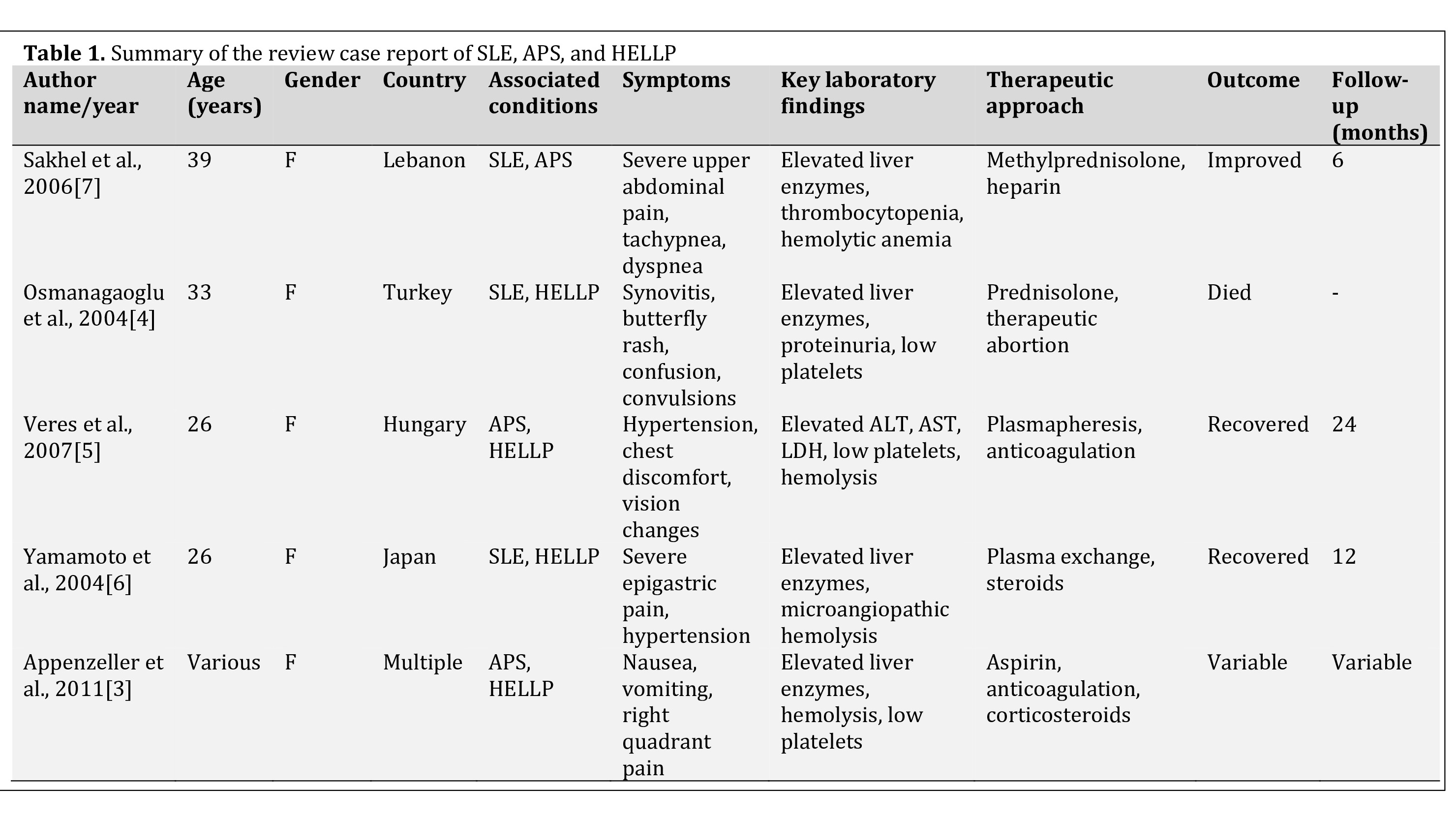
Three in One: Systemic Lupus Erythematosus, HELLP Syndrome, and Antiphospholipid Syndrome: A Case Report and Literature Review
Sivan H. Salih, Dana T. Gharib, Nahida H. Ahmad, Hoshmand R. Asaad, Ronak S. Ahmed, Huda M....
Abstract
Introduction
Systemic lupus erythematosus (SLE) is a multisystem autoimmune disease commonly affecting women of reproductive age. Its overlap with HELLP syndrome (Hemolysis, Elevated Liver Enzymes, and Low Platelet Count) and antiphospholipid syndrome (APS) during pregnancy is rare and presents diagnostic and therapeutic challenges due to shared clinical and laboratory features. This report describes a case of pre-existing SLE complicated by the concurrent occurrence of SLE flare, HELLP syndrome, and APS during pregnancy.
Case presentation
A 26-year-old female with known SLE presented with right upper quadrant abdominal pain and hypertension two days following a mid-trimester abortion at 22 weeks of gestation. Laboratory evaluation revealed thrombocytopenia, elevated liver enzymes, and hemolysis, consistent with HELLP syndrome. The presence of lupus anticoagulant and anticardiolipin antibodies, in conjunction with her obstetric history, supported the diagnosis of APS. The patient was treated with high-dose corticosteroids and anticoagulation, resulting in significant clinical improvement.
Literature review
A review of literature highlights the consistent presentation of nonspecific symptoms such as abdominal pain, nausea, and hypertension in patients with overlapping SLE, HELLP syndrome, and APS. Laboratory findings often reveal thrombocytopenia, elevated liver enzymes, and hemolysis, reflective of underlying microangiopathic processes. Therapeutic strategies typically involve corticosteroids and anticoagulation, with plasmapheresis reserved for severe cases.
Conclusion
The coexistence of SLE flare, HELLP syndrome, and APS during pregnancy is a rare and complex condition that requires careful evaluation. Early recognition and appropriate management are crucial for achieving favorable outcomes.
Introduction
Systemic lupus erythematosus (SLE) is a chronic autoimmune disease with multisystem involvement, affecting approximately 20 to 150 per 100,000 individuals worldwide, with a strong female predominance [1,2]. It commonly presents with fatigue, arthralgia, skin manifestations, and renal or hematological complications, but its presentation can vary widely depending on disease activity [3]. Pregnancy in patients with SLE is considered high-risk due to increased risks of preeclampsia, fetal loss, and preterm delivery [4]. Antiphospholipid syndrome (APS) is another autoimmune disorder that frequently overlaps with SLE. It is characterized by the presence of antiphospholipid antibodies and is associated with thrombotic events and pregnancy complications such as recurrent miscarriage and intrauterine growth restriction [5,6]. HELLP (Hemolysis, Elevated Liver Enzymes, and Low Platelet Count) syndrome is a severe form of preeclampsia that can develop in up to 0.9% of pregnancies, usually leading to hepatic dysfunction, hypertension, and coagulopathy [7]. These conditions exhibit overlapping clinical and laboratory features such as thrombocytopenia, elevated liver enzymes, and hemolysis, making diagnosis and management particularly challenging [3-7].
This report presents a known case of SLE with the simultaneous occurrence of SLE flare, HELLP syndrome, and APS during pregnancy. The report was prepared in accordance with the CaReL guidelines, with all references evaluated for reliability and scientific integrity [8,9].
Case Presentation
Patient information
A 26-year-old woman presented with a one-week history of right upper quadrant abdominal pain. She had a second-trimester abortion at 22 weeks of gestation, delivered vaginally two days earlier. Her medical history included SLE, diagnosed eight years earlier following a miscarriage. Because of recurrent abortions, she was placed on aspirin during pregnancy. She also had a history of gestational hypertension.
Clinical findings
Examination revealed significant tenderness in the epigastric region upon light palpation, with no changes in mental status.
Diagnostic approach
Laboratory investigations revealed markedly elevated liver enzymes, including alanine aminotransferase (ALT: 2500 U/L; reference: 7–56 U/L) and aspartate aminotransferase (AST: 3000 U/L; reference: 10–40 U/L). She also had thrombocytopenia, with a platelet count of 50,000/mm³ (reference: 150,000–450,000/mm³), and a prolonged prothrombin time (PT) of 22 seconds (reference: 11–15 seconds) with an elevated international normalized ratio (INR: 2.0; reference: 0.8–1.2). D-dimer levels were markedly elevated at 2500 ng/mL (reference: <500 ng/mL), while fibrinogen was decreased to 50 mg/dL (reference: 200–400 mg/dL). Peripheral blood smear demonstrated schistocytes, indicative of ongoing microangiopathy. Autoimmune testing confirmed active SLE, with a positive antinuclear antibody (ANA) and elevated anti-double-stranded DNA (anti-dsDNA) titers. Imaging studies, including an abdominal computed tomography scan, demonstrated severe hepatic necrosis without evidence of rupture or hematoma. These findings confirmed the coexistence of SLE flare, HELLP syndrome, and APS, a rare and complex presentation.
Therapeutic intervention and follow-up
The patient was admitted to the intensive care unit for supportive care and initiated methylprednisolone pulse therapy for three consecutive days, resulting in significant improvement. Liver enzymes normalized, platelet counts increased, and symptoms resolved. Anticoagulation with warfarin was started for APS, and hydroxychloroquine was continued for SLE management. At discharge, she was stable and referred to rheumatology for long-term follow-up.
Discussion
Pregnancy in patients with SLE carries an increased risk of adverse maternal and fetal outcomes, including preeclampsia, fetal loss, and preterm birth, particularly in cases complicated by active disease [5]. The presence of APS, a thrombotic disorder defined by antiphospholipid antibodies, further increases pregnancy risks by promoting thrombosis, recurrent miscarriages, and placental insufficiency [6].
HELLP syndrome, a severe form of preeclampsia, presents with hepatic dysfunction, endothelial injury, and hematologic abnormalities, leading to substantial maternal and fetal morbidity [7].
The present case developed HELLP syndrome and APS following a mid-trimester abortion at 22 weeks, reflecting the complex interplay of these conditions and the challenges in diagnosis and management. A brief literature review identified 4 reported cases of concurrent (SLE and APS), (SLE and HELLP syndrome), or (APS and HELLP syndrome). All occurred in young women aged 26–39 years, underscoring the well-established female predominance of these conditions (Table 1) [3-7].
Author name/year |
Age (years) |
Gender |
Country |
Associated conditions |
Symptoms |
Key laboratory findings |
Therapeutic approach |
Outcome |
Follow-up (months) |
Osmanagaoglu et al., 2004[4] |
33 |
F |
Turkey |
SLE, HELLP |
Synovitis, butterfly rash, confusion, convulsions
|
Elevated liver enzymes, proteinuria, and low platelets |
Prednisolone, therapeutic abortion |
Died |
- |
Veres et al., 2007[5] |
26 |
F |
Hungary |
APS, HELLP |
Hypertension, chest discomfort, vision changes
|
Elevated ALT, AST, LDH, low platelets, hemolysis |
Plasmapheresis, anticoagulation |
Recovered |
24 |
Yamamoto et al., 2004[6] |
26 |
F |
Japan |
SLE, HELLP |
Severe epigastric pain, hypertension
|
Elevated liver enzymes, microangiopathic hemolysis |
Plasma exchange, steroids |
Recovered |
12 |
Sakhel et al., 2006[7] |
39 |
F |
Lebanon |
SLE, APS |
Severe upper abdominal pain, tachypnea, dyspnea
|
Elevated liver enzymes, thrombocytopenia, hemolytic anemia |
Methylprednisolone, heparin |
Improved |
6 |
SLE: Systemic Lupus Erythematosus; APS: Antiphospholipid Syndrome; HELLP: Hemolysis, Elevated Liver enzymes, Low Platelets; F: Female; ALT: Alanine Aminotransferase; AST: Aspartate Aminotransferase; LDH: Lactate Dehydrogenase |
Pregnancy is a known trigger for SLE flares, and the additional presence of APS further increases the risk of obstetric complications [2,4,5]. The present case is consistent with these observations, as the patient’s history of SLE and pregnancy loss conferred an elevated risk for the development of APS and HELLP syndrome.
The most common symptoms reported in the reviewed cases were right upper quadrant abdominal pain, hypertension, nausea, and dyspnea, which are classic signs of HELLP syndrome [3-7]. Sakhel et al. reported tachypnea and dyspnea, which can indicate pulmonary involvement or preeclampsia-related respiratory distress [7]. Osmanagaoglu et al. reported another case presenting with synovitis, butterfly rash, and confusion, indicative of either neurological lupus involvement or eclampsia [4]. Some cases presented with vision disturbances and chest discomfort, which may indicate APS-related microvascular thrombotic events [5]. While some patients had neurological involvement, the present case exhibited a severe hypertensive episode, reinforcing the complexity of these overlapping conditions.
Laboratory investigations across cases demonstrated consistent hematologic abnormalities, including thrombocytopenia, elevated liver enzymes, and hemolysis, which are defining features of HELLP syndrome [3]. One case reported proteinuria, which is more suggestive of SLE nephritis or preeclampsia-related renal dysfunction [4]. In another case, hemolysis was confirmed through microangiopathic findings rather than an immune-mediated mechanism [6]. Patients with APS frequently exhibited prolonged PT/INR, elevated D-dimer levels, and thrombocytopenia, reflecting the hypercoagulable state of APS [5].
The present case demonstrated severe thrombocytopenia, elevated ALT/AST, and hemolysis, with positive lupus anticoagulant and anticardiolipin antibodies confirming APS and elevated anti-dsDNA titers indicating active SLE. This profile closely mirrors the reviewed cases of the literature, emphasizing the diagnostic overlap between HELLP syndrome, SLE flares, and APS.
Although imaging findings were not reported in all cases, hepatic involvement was a significant feature in those with severe HELLP syndrome or APS-associated thrombosis. Osmanagaoglu et al. reported a case of hepatic infarctions and subcapsular hematomas, suggesting vascular damage secondary to HELLP syndrome or APS-related thrombosis [7]. Veres et al. described another case with hepatic microangiopathic changes in the absence of infarction, highlighting the diverse spectrum of hepatic involvement in such patients [5]. The present case exhibited hepatic involvement in imaging, consistent with hepatic dysfunction in severe HELLP syndrome, reinforcing the value of imaging studies in distinguishing these conditions.
Management strategies varied depending on the severity of the disease, but high-dose corticosteroids were universally used to suppress SLE flares and reduce systemic inflammation [6]. APS patients received anticoagulation therapy with heparin or warfarin to prevent thrombotic complications. In severe cases, plasmapheresis and intravenous immunoglobulin were administered to manage catastrophic APS or refractory hemolysis [5]. The case reported by Osmanagaoglu et al. required therapeutic abortion due to the severity of maternal complications, highlighting the need for individualized obstetric decision-making in these high-risk pregnancies [4].
The present case was treated with methylprednisolone for SLE flare and heparin for APS, resulting in significant clinical improvement, which mirrors the treatment success observed in the reviewed cases (Table 1).
Outcomes among reported cases varied based on disease severity and timing of intervention. While most patients responded well to treatment, the case reported by Osmanagaoglu et al. resulted in maternal mortality, particularly due to multi-organ failure and delayed diagnosis [4]. Other cases reported full recovery following corticosteroid therapy and anticoagulation [5,6]. The case reported by Appenzeller et al. demonstrated partial resolution of symptoms but continued to require long-term monitoring due to APS-related complications [3].
Early intervention played a crucial role in improving prognosis, as seen in cases that received timely corticosteroid and anticoagulation therapy. The present case, diagnosed early and treated promptly, achieved clinical stabilization, underscoring the critical role of timely diagnosis and multidisciplinary management in improving maternal outcomes.
Conclusion
The coexistence of SLE flare, HELLP syndrome, and APS during pregnancy is a rare and complex condition that requires careful evaluation. Early recognition and appropriate management are crucial for achieving favorable outcomes.
Declarations
Conflicts of interest: The authors declare no conflicts of interest.
Ethical approval: Not applicable.
Patient consent (participation and publication): Written informed consent was obtained from the patient for participation and publication.
Funding: The present study received no financial support.
Acknowledgements: None to be declared.
Authors' contributions: SHS and RSA were significant contributors to the conception of the study and the literature search for related studies. KAN and HAN were involved in the literature review, study design, and manuscript writing. DTG, NHA, HRA, HMM, JIH, and KFH were involved in the literature review, study design, critical revision of the manuscript, and table processing. RSA and HAN confirm the authenticity of all the raw data. All authors approved the final version of the manuscript.
Use of AI: ChatGPT-4 was used to assist in language editing and improving the clarity of the manuscript. All content was reviewed and verified by the authors. Authors are fully responsible for the entire content of their manuscript.
Data availability statement: Not applicable.